
Подборка снимков экрана (скриншотов) программы
Путь: начало » скриншоты » стр. 1
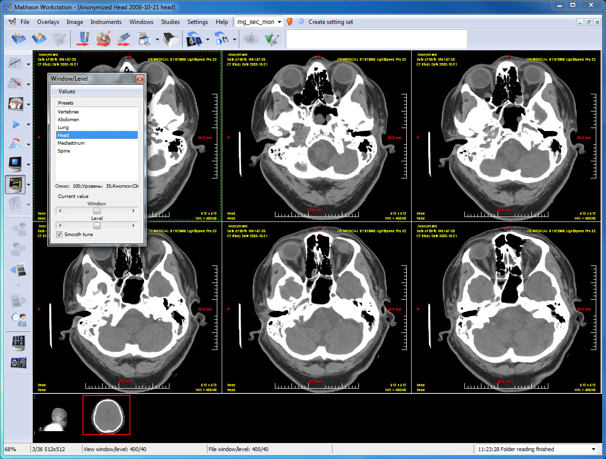
2x3 splitting, CT series, one of the pre-set 'window/level' value is applied.
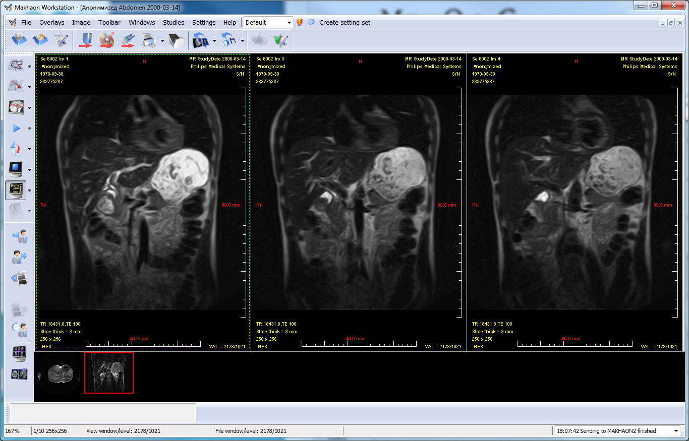
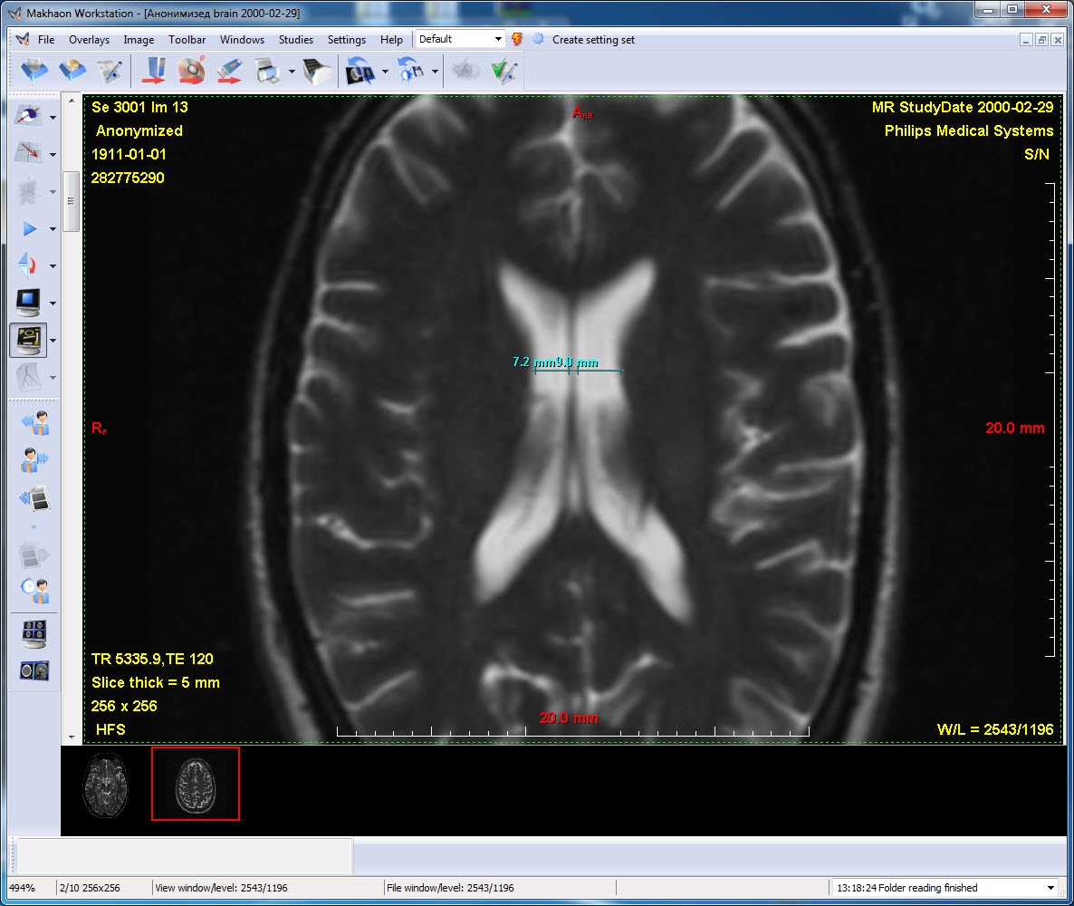
1x3 splitting, MR series
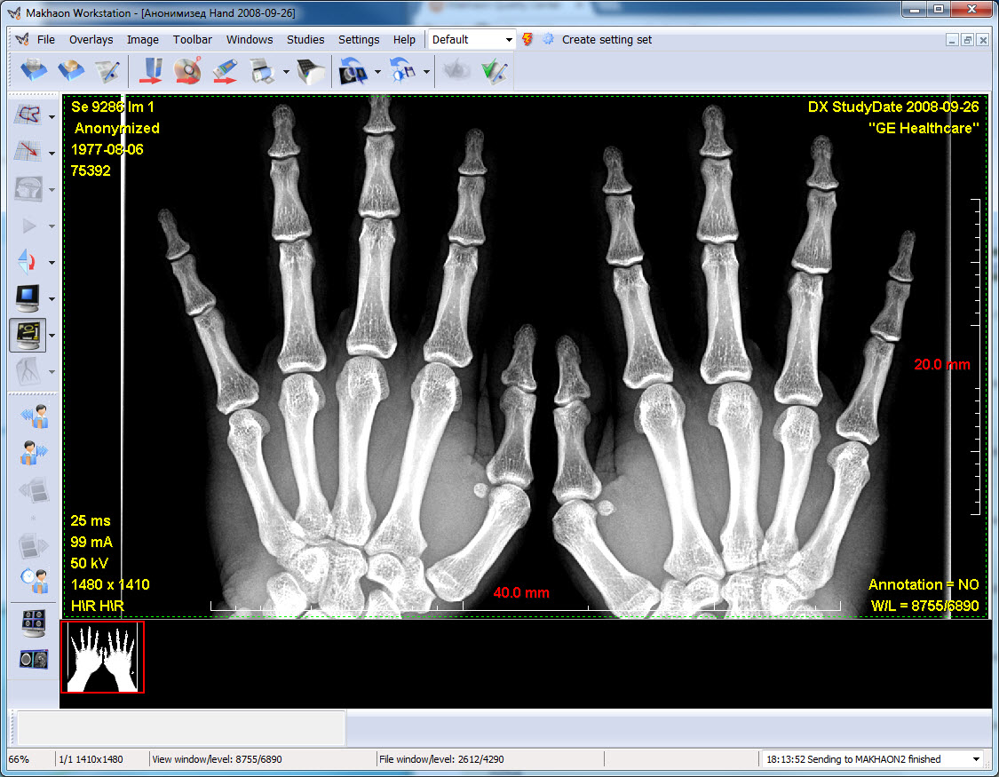
1x1 splitting, DX series
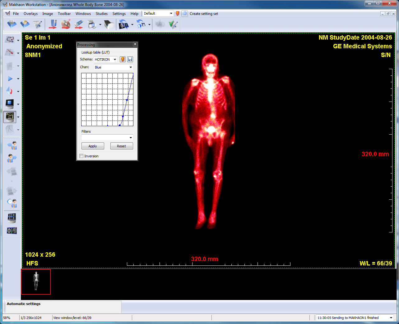
Compressed NM-image, Hot Iron color LUT is applied.
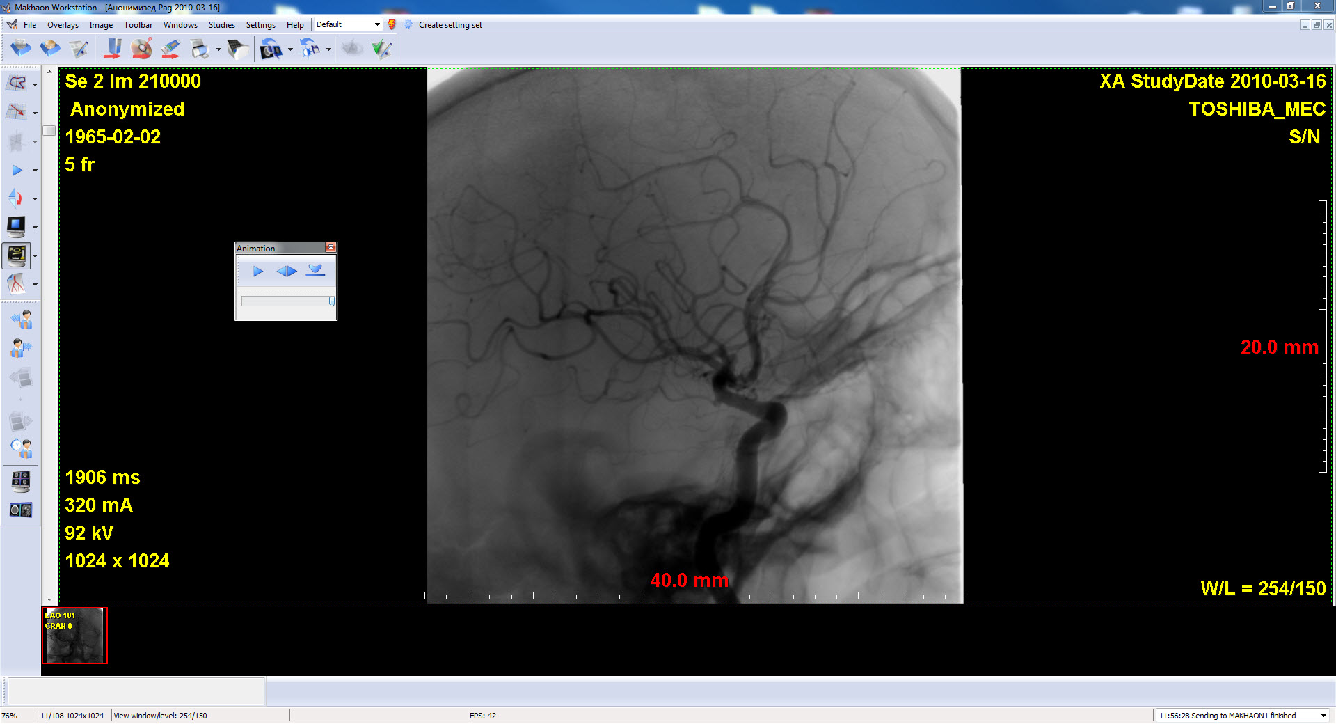
XA-series animation, 1024x1024 series resolution, 1680x1050 display resolution, maximum speed is 41 frames a second. Speed, enough for the real time mode is 25 frames a second, real time mode is disabled.
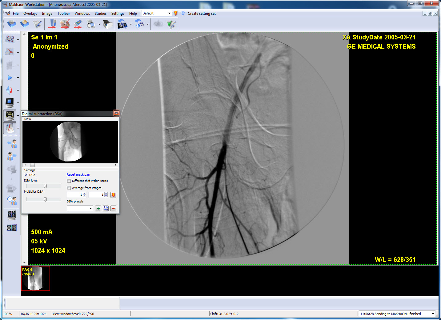
XA-series. Digital subtraction angiography mode (DSA-mode). The mask is slightly offset comparing to the image position. The mask number is switched automatically for every image (on the basis of DICOM-headline information).
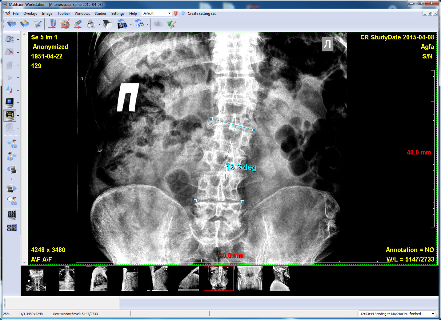
CR series. Cobb angle measurements.
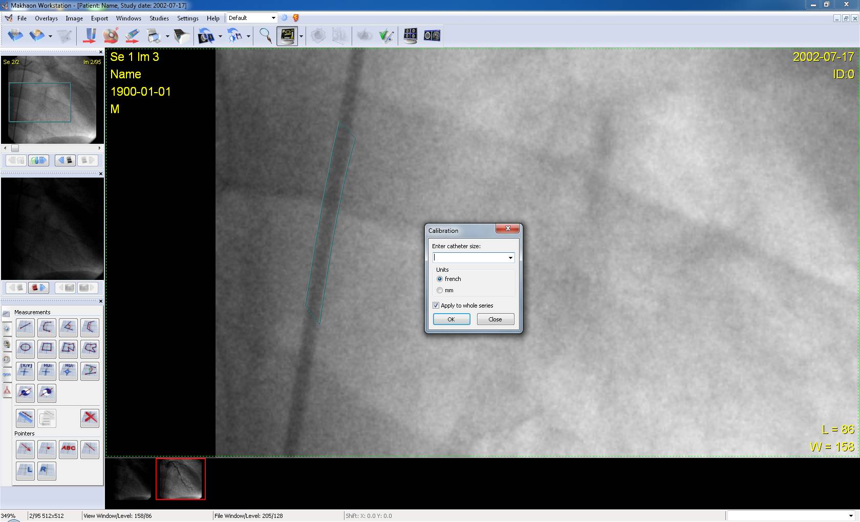
XA-series. The automatic image calibration or series calibration on the catheter caliber specified.
Automatic sizing of the image light areas. The "Light gradient segmentclear gradient section" tool.
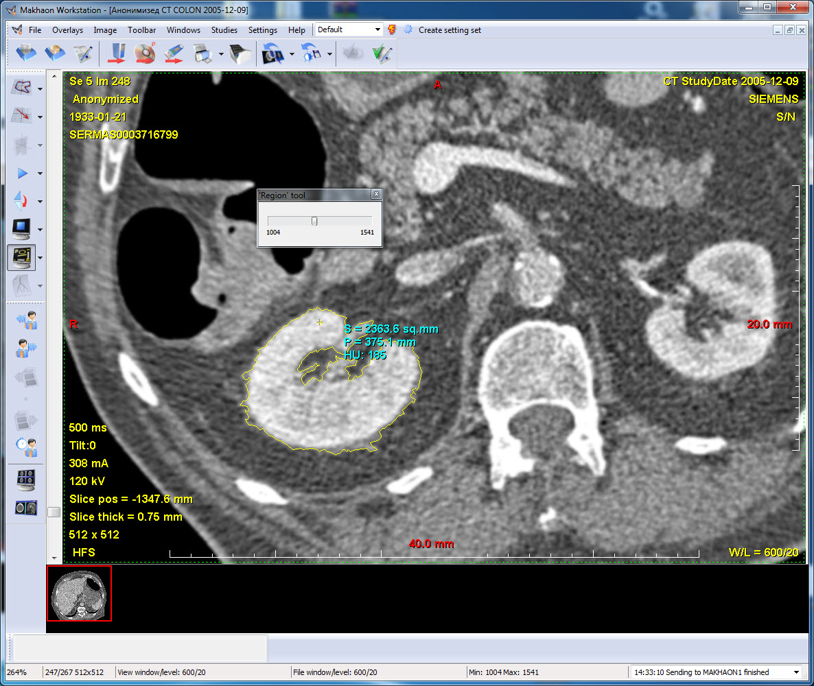
Automatic image contrasting region search. The "Region" tool. The result is the overlay equivalent to the "hands-free" tool. Area and perimeter calculation, calculation of HU-overlay value averaged through all the area.
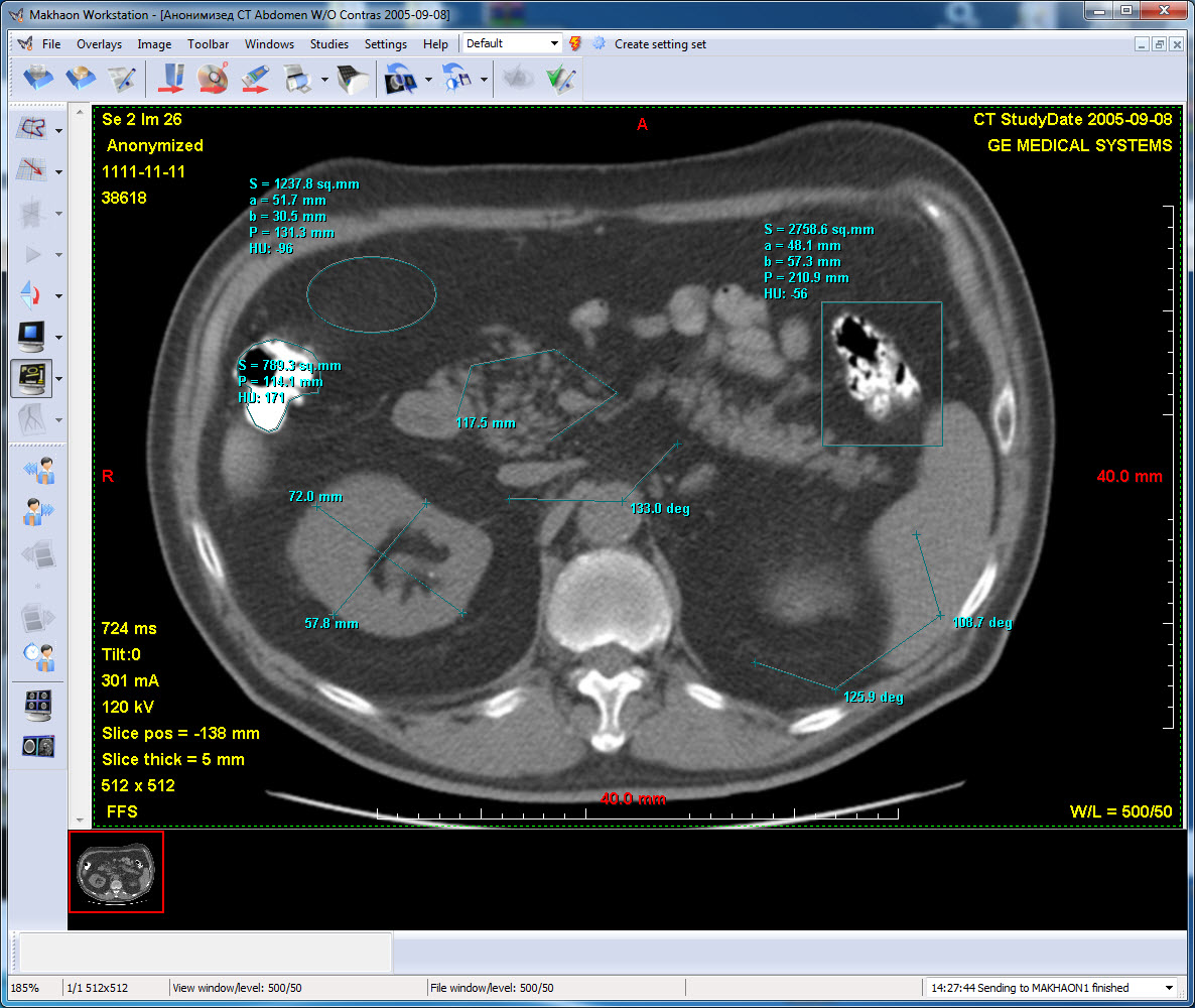
CT series. Different overlays.
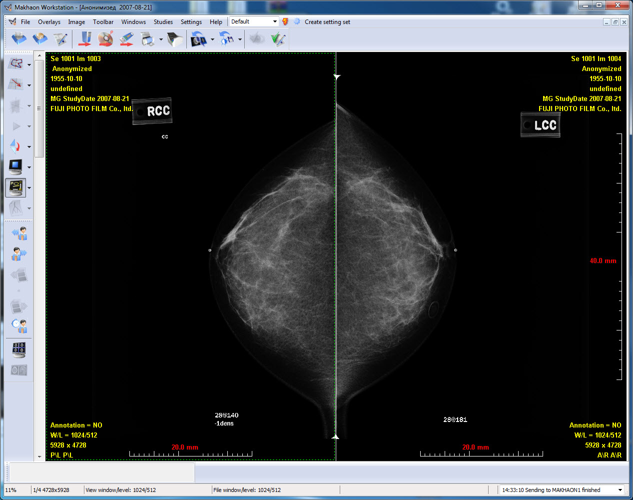
MG series. The mammograms display mode is switched automatically on one of the displays (there is one display in the system).
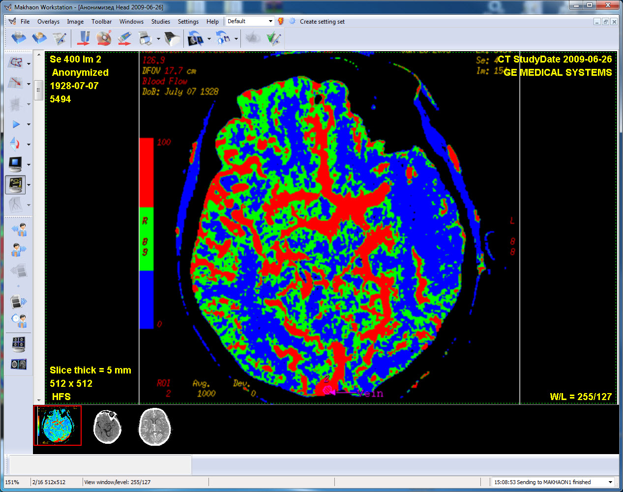
CT series. Diffusion calculation result of the CT images.
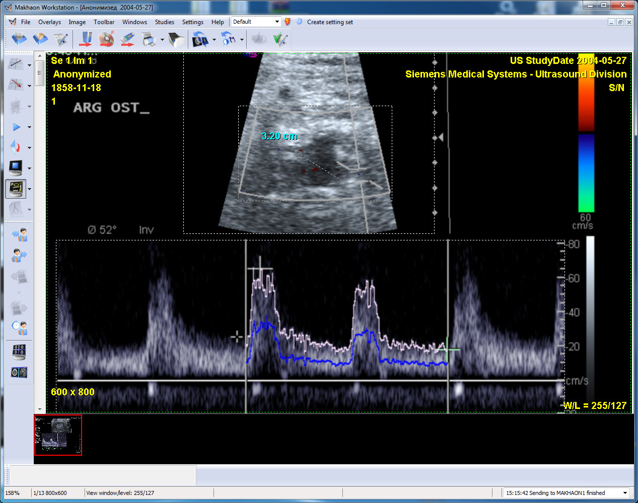
US series. Calibrated regions are marked. Sizing is applied on one of the areas.

US series. Several measurements are made in different calibration regions. If the measurements in the low area is parallel to one of the coordinate axis, then the section length is calibrated according to the corresponding coordinate. If the section is inclined, then the length is calculated for two coordinates at one and the same time.
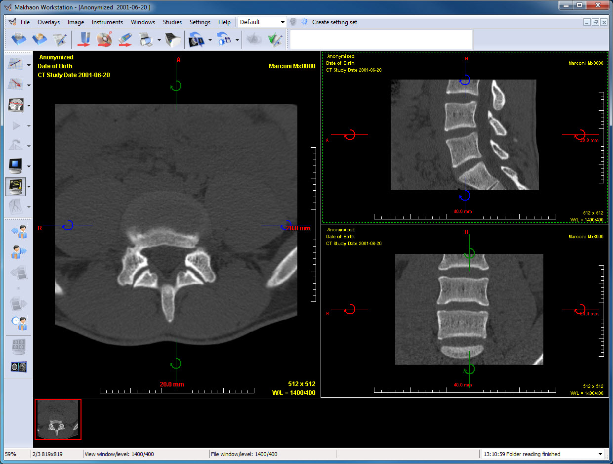
CT series. The multiplanar reconstruction mode (MPR).
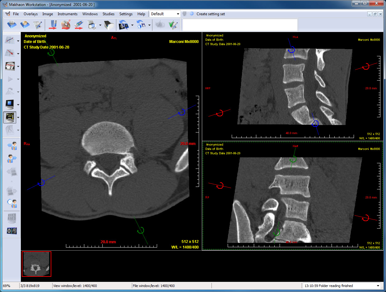
CT series. The double oblique multiplanar reconstruction mode (Double obliq MPR).
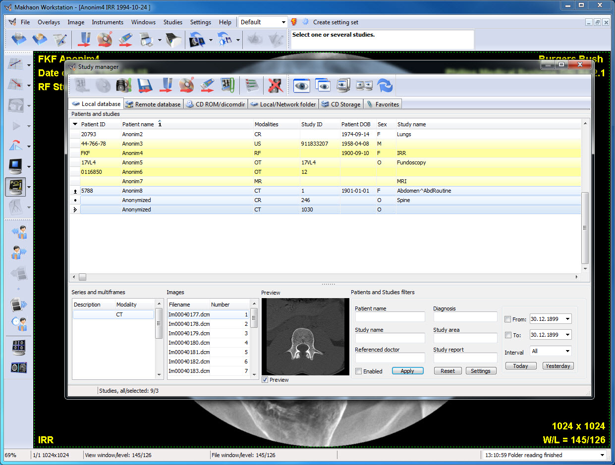
Study manager. There are three studies chosen for simultaneous opening and view.
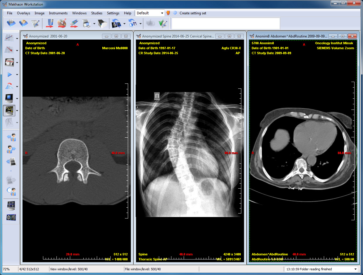
Three studies are opened simultaneously.
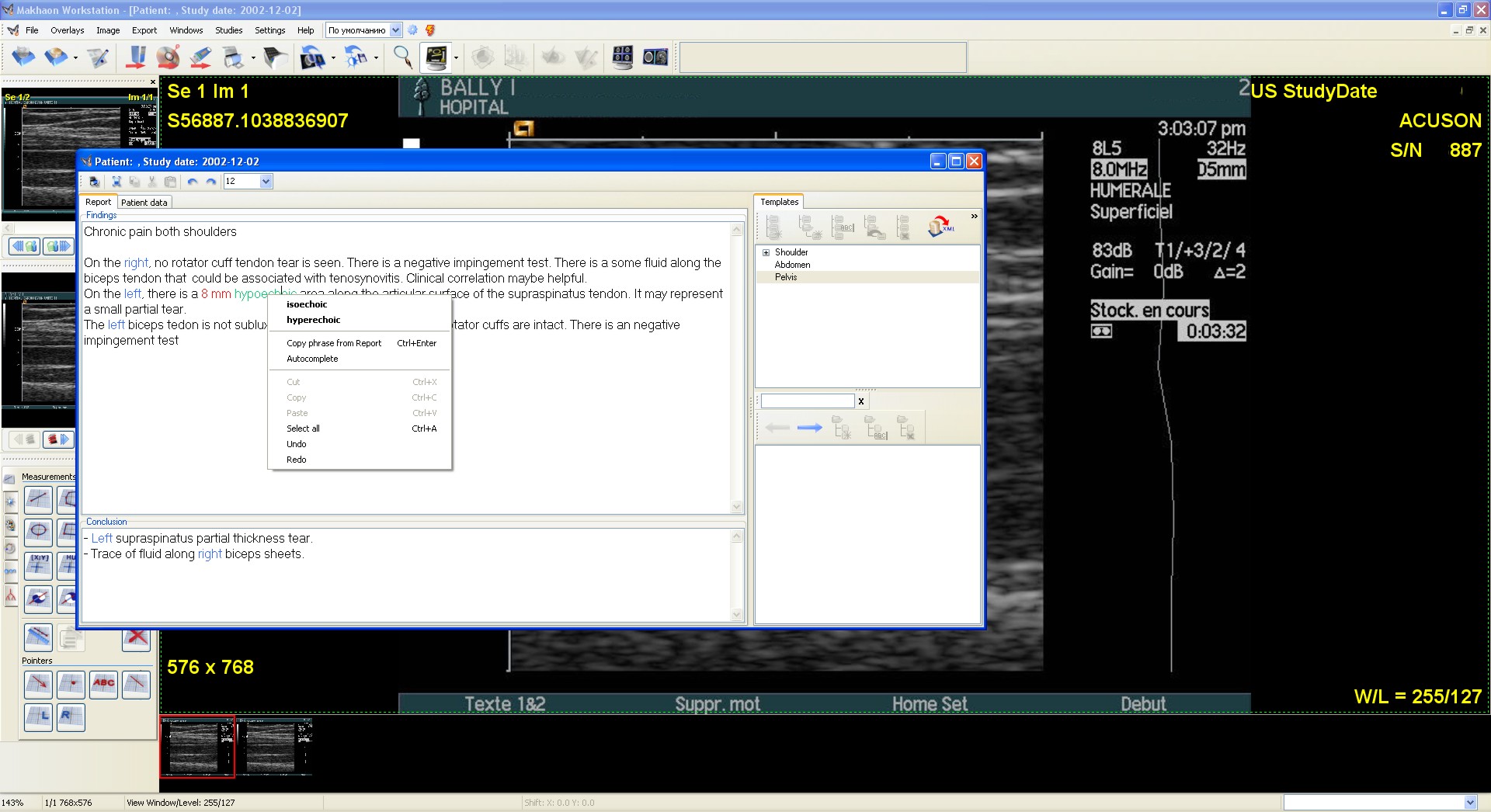
Patient's study description window. There are report, medical conclusion, templates; the context menu includes the phrase-substitute variants (phrase substitution is set in the Configurator).
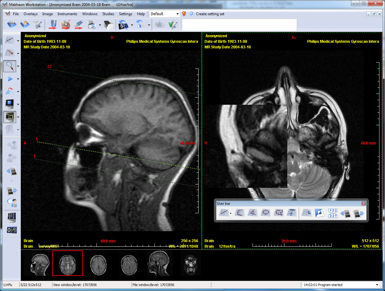
The MR study is opened, two series are opened simultaneously, the screen magnifier mode is activated, and user's panel is displayed. The user's panel (as well as the other tools bars can be set at your convenience and placed at any side of your software interface).
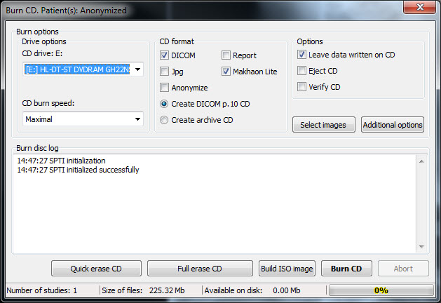
The optical disk-write window.
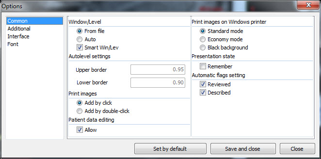
Program settings. Tab "Common".

Program settings. Tab "Additional parameters".
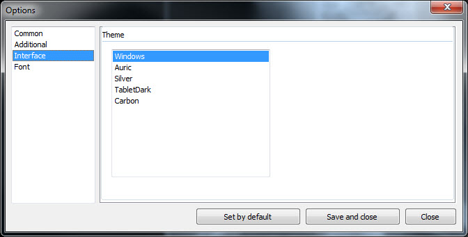
Program settings. Tab "Interface". The Office 2003 interface scheme is activated.
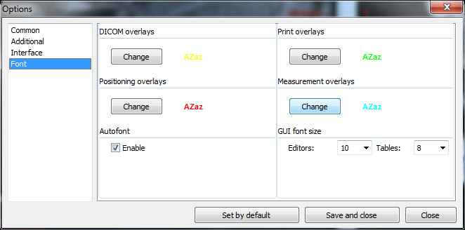
Program settings. Tab "Font".
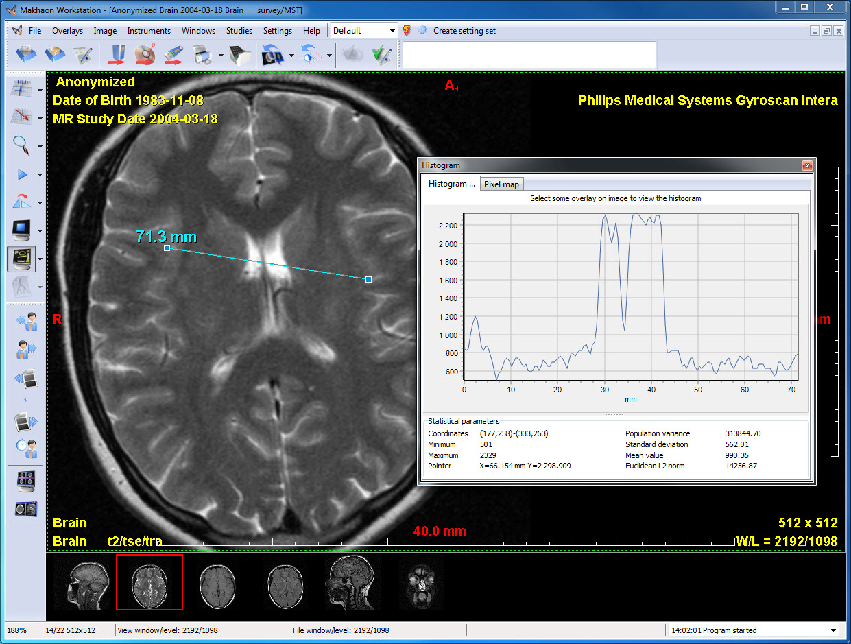
"Histogram" window. HU-values histogram is displayed for the section chosen. The cursor is on the histogram (it is cursored with the PC mouse as you point it on the histogram window). The HU value at the cursor point is displayed in the window: Cursor X=102.844 mm Y = 2009.273. The X-axis values can be calibrated on their length (in mm) - in case the image is calibrated.
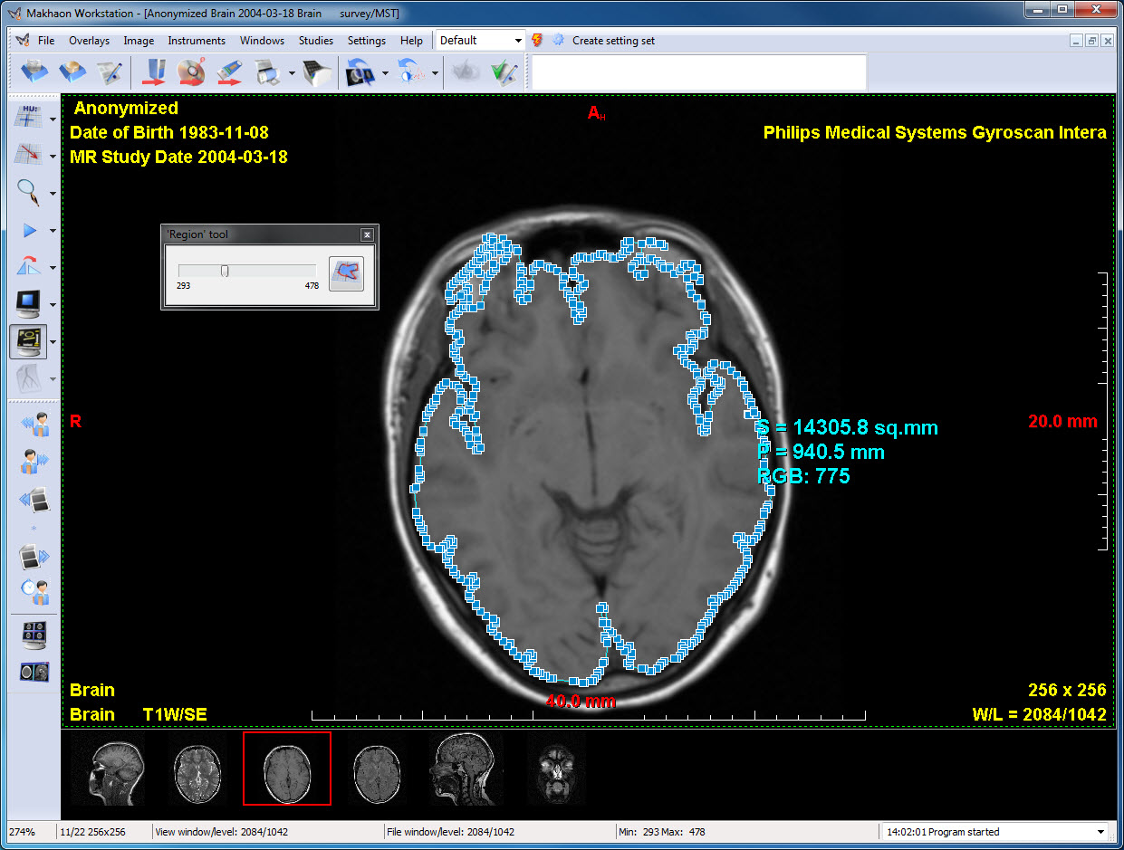
"Region" tool. Image contrast region search.

"Makhaon configurator" software. Tab "Database".
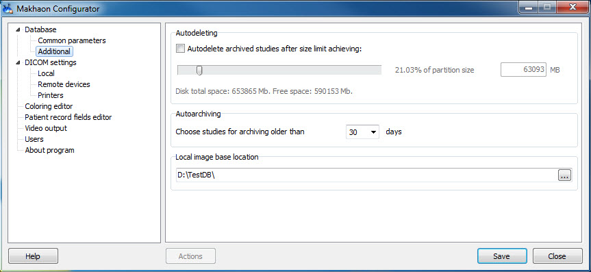
"Makhaon configurator" software. Tab "Additional parameters".
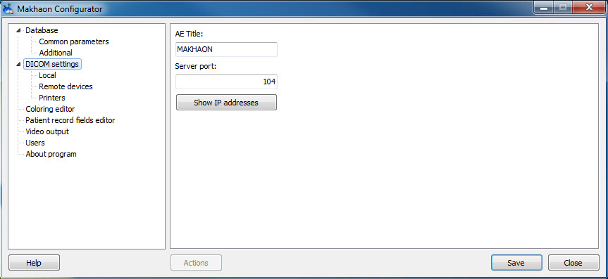
"Makhaon configurator" software. Tab "DICOM parameters".
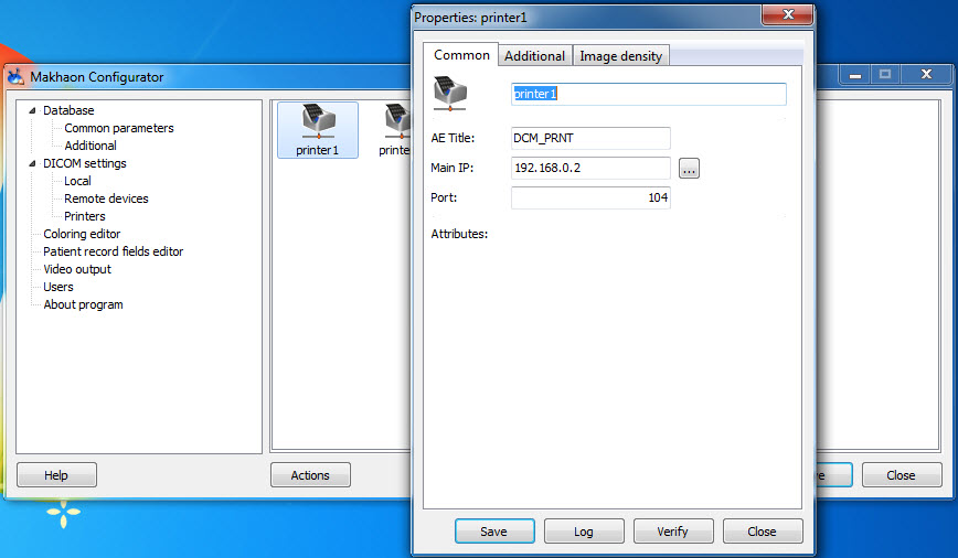
"Makhaon configurator" software. Tab "Printers".
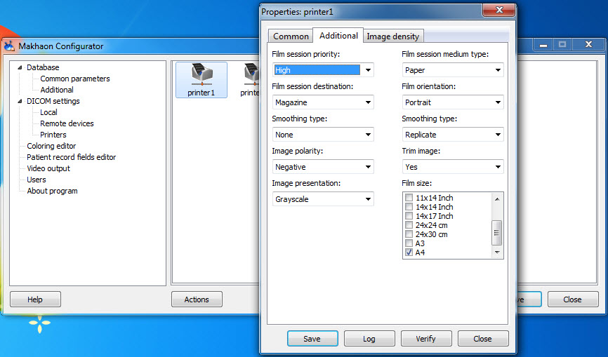
"Makhaon configurator" software. Tab "Printers".
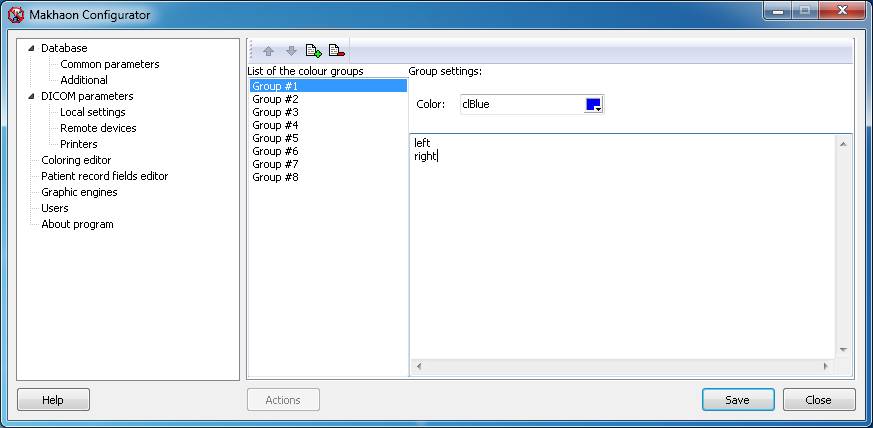
"Makhaon configurator" software. Tab "Coloring editor".
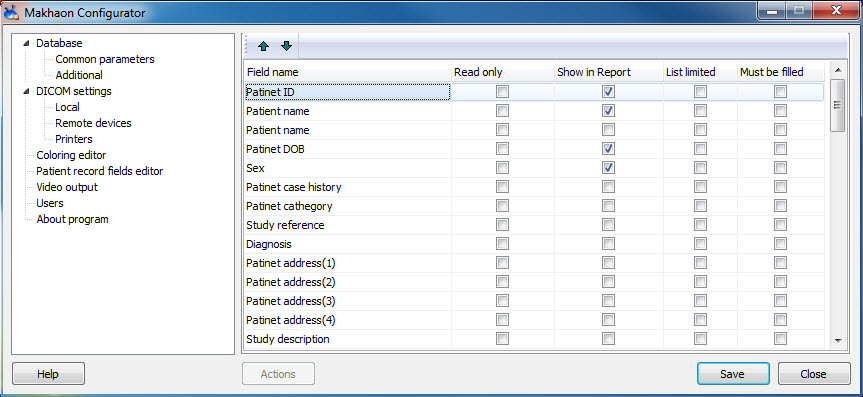
"Makhaon configurator" software. Tab "Patient info fields editor".
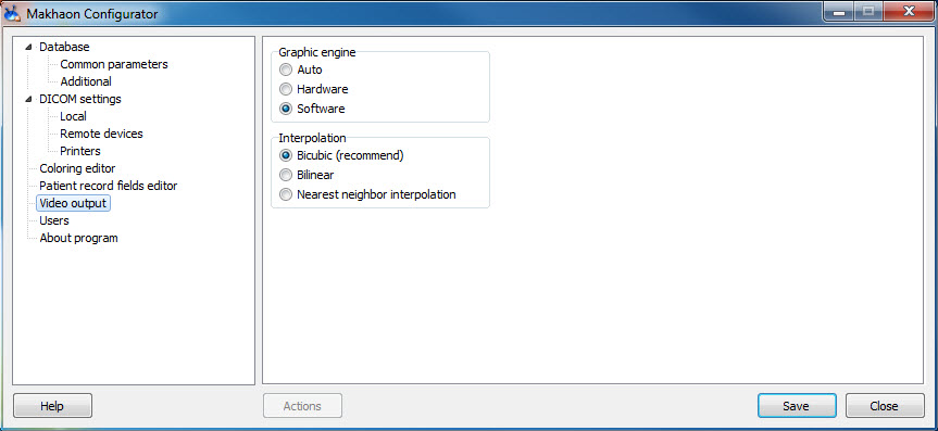
"Makhaon configurator" software. Tab "Graphics display'.
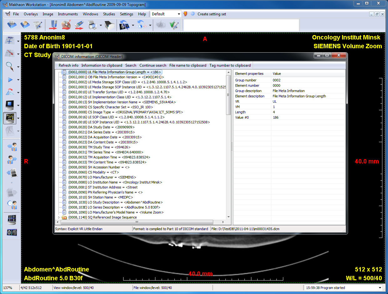
Information window about DICOM image.
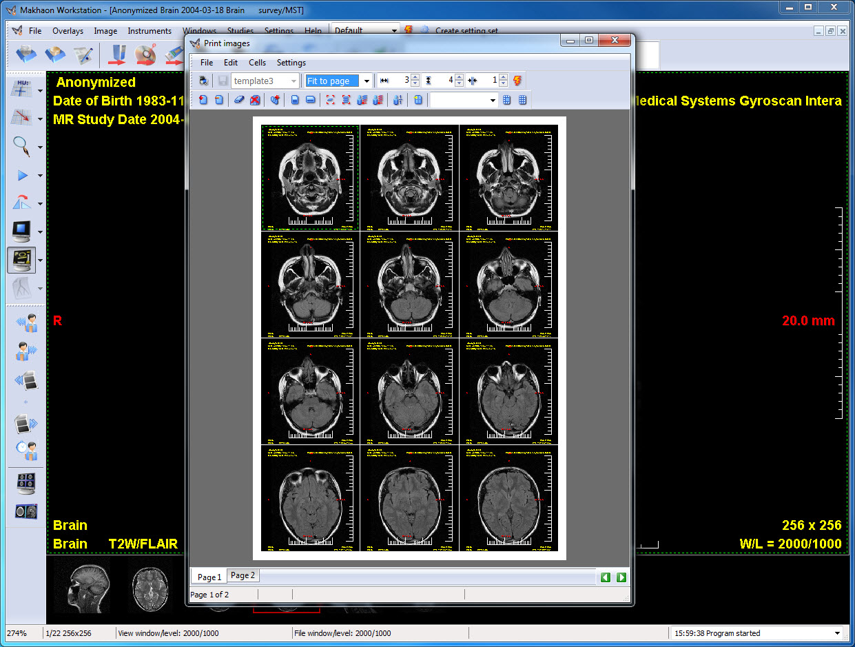
Print image window. There are several images and scanogram added for print. Three images are chosen and the intersection lines of scanogram are formed for these three images. ("Assign scanogam" tool).
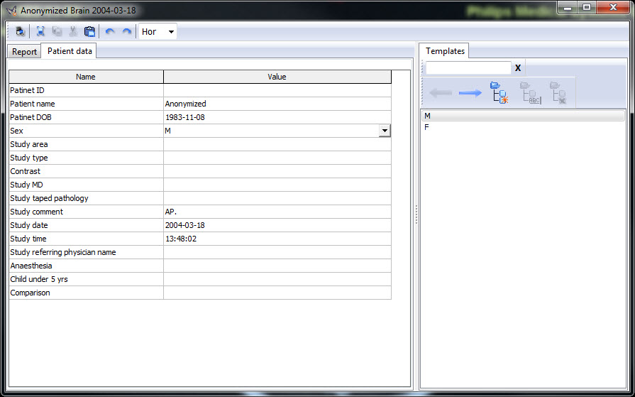
Study report window, patient information. The study and patient data and templates are displayed.
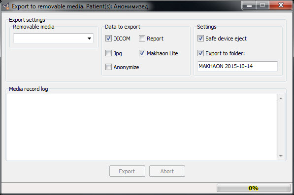
Window of store studies to the removable media (to the flash disk).
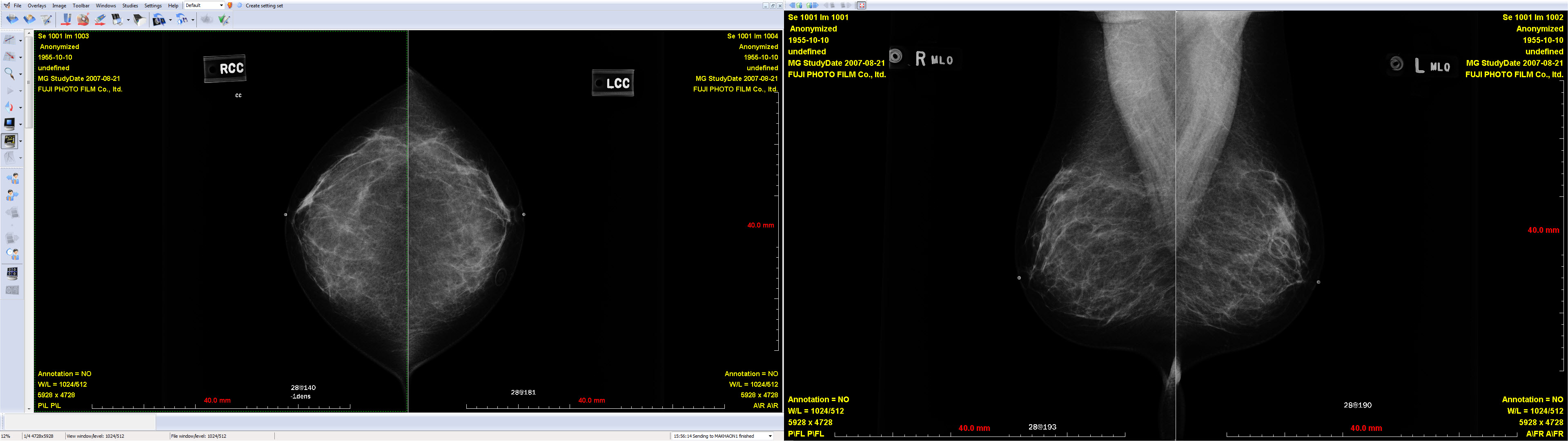
MG-series. The mammogram display mode is automatically activated. Program interface is set optimally for such images view (on two displays). All the needed tools can be added to the bar.
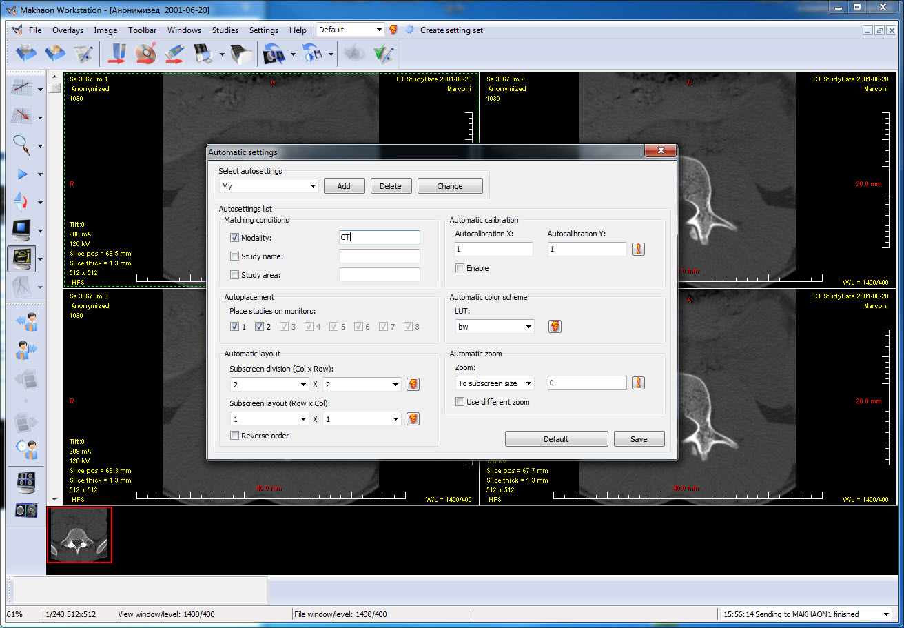
"Automatic settings" window. For the main window "CT" setting is applied. For all of CT series the 2x2 splitting will be applied automatically. For all the other series splitting will be on default. At the present time there is 2x2 splitting.
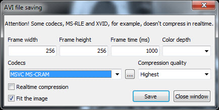
The AVI-file series store window.
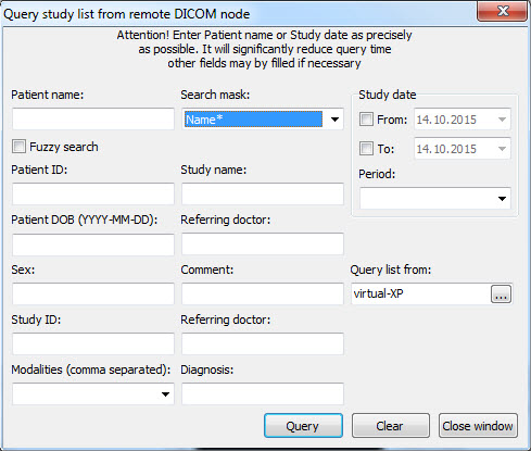
The remote device studies list enquiry window (DICOM Query).
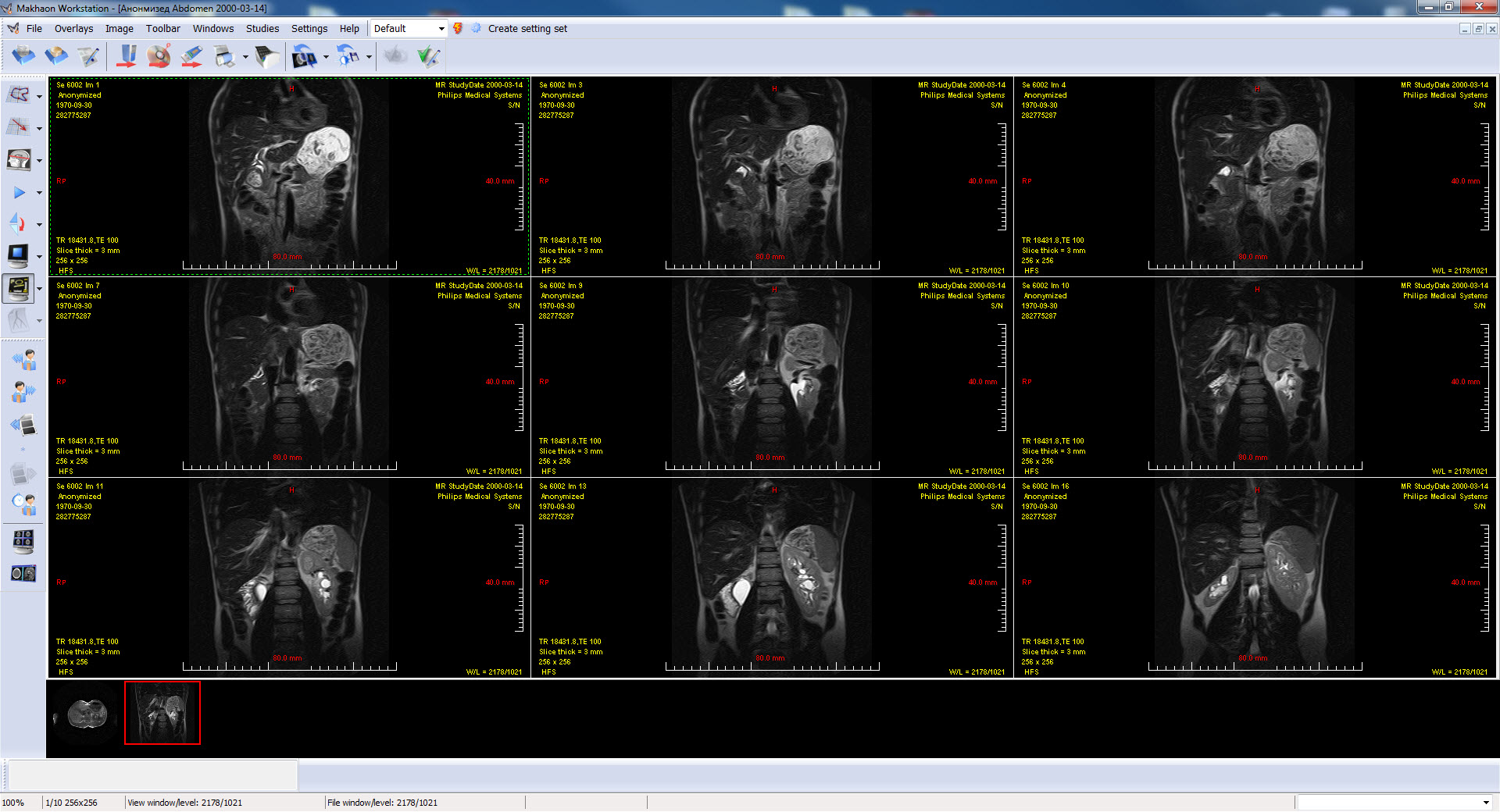
The ordinary program interface, 3x3 splitting, MR series.