
Подборка снимков экрана (скриншотов) программы
Путь: начало » скриншоты » стр. 2
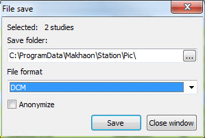
Different formats files storage on a disk window. DICOM format is chosen, the anonymization is activated.

The main program window bar setting window. Tab "Toolbars".
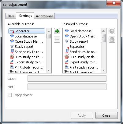
The main program window bar setting window. Tab "Settings".
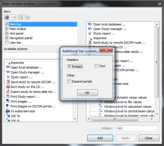
The main program window bar setting window. Tab "Additional".
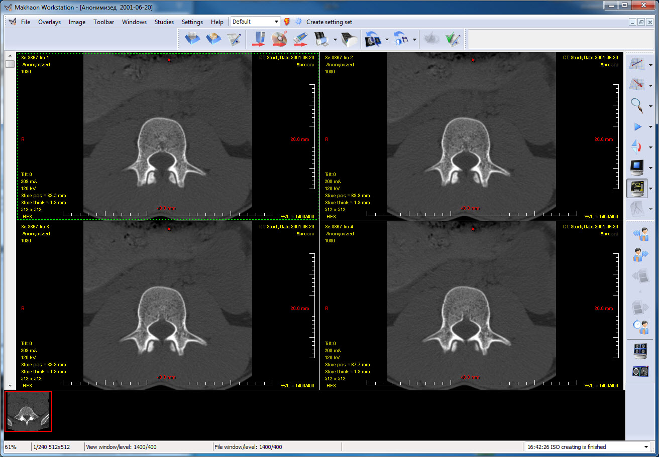
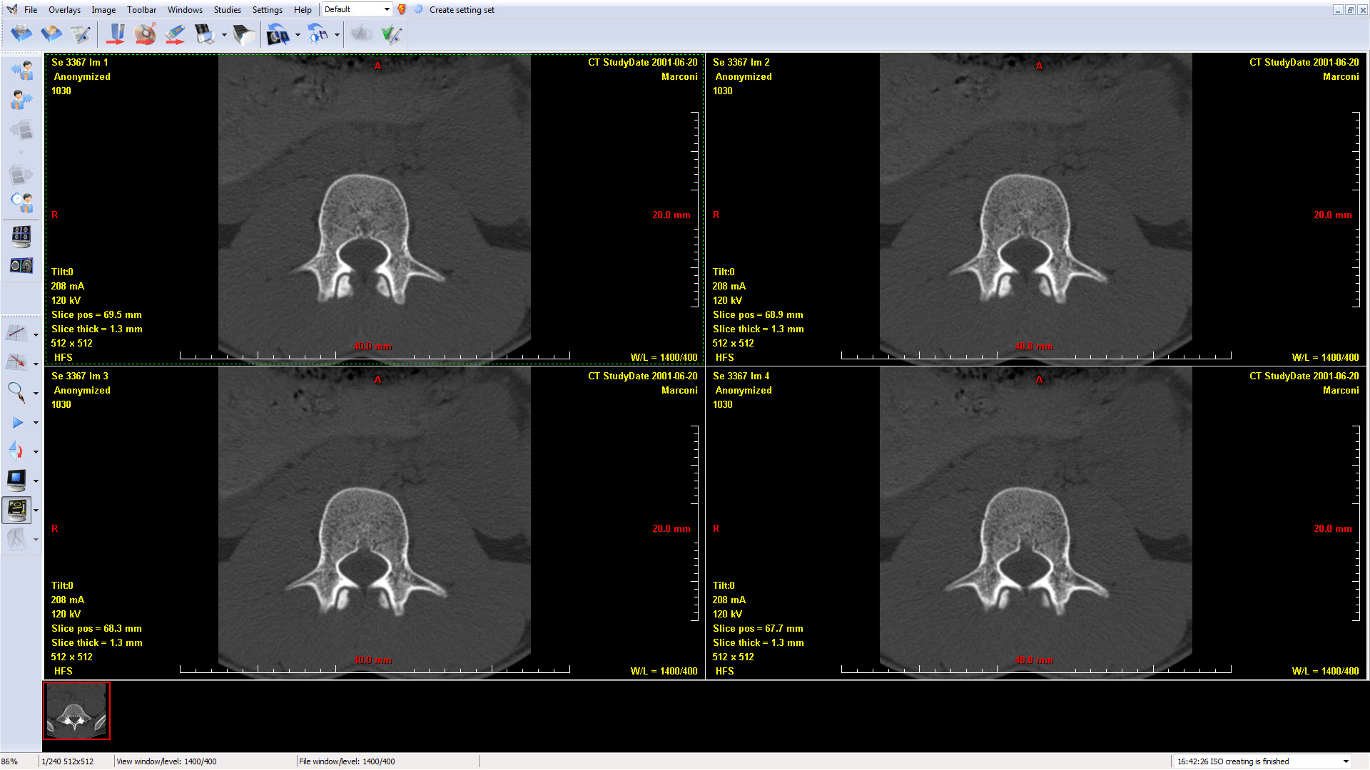
Main window. Controls position option.
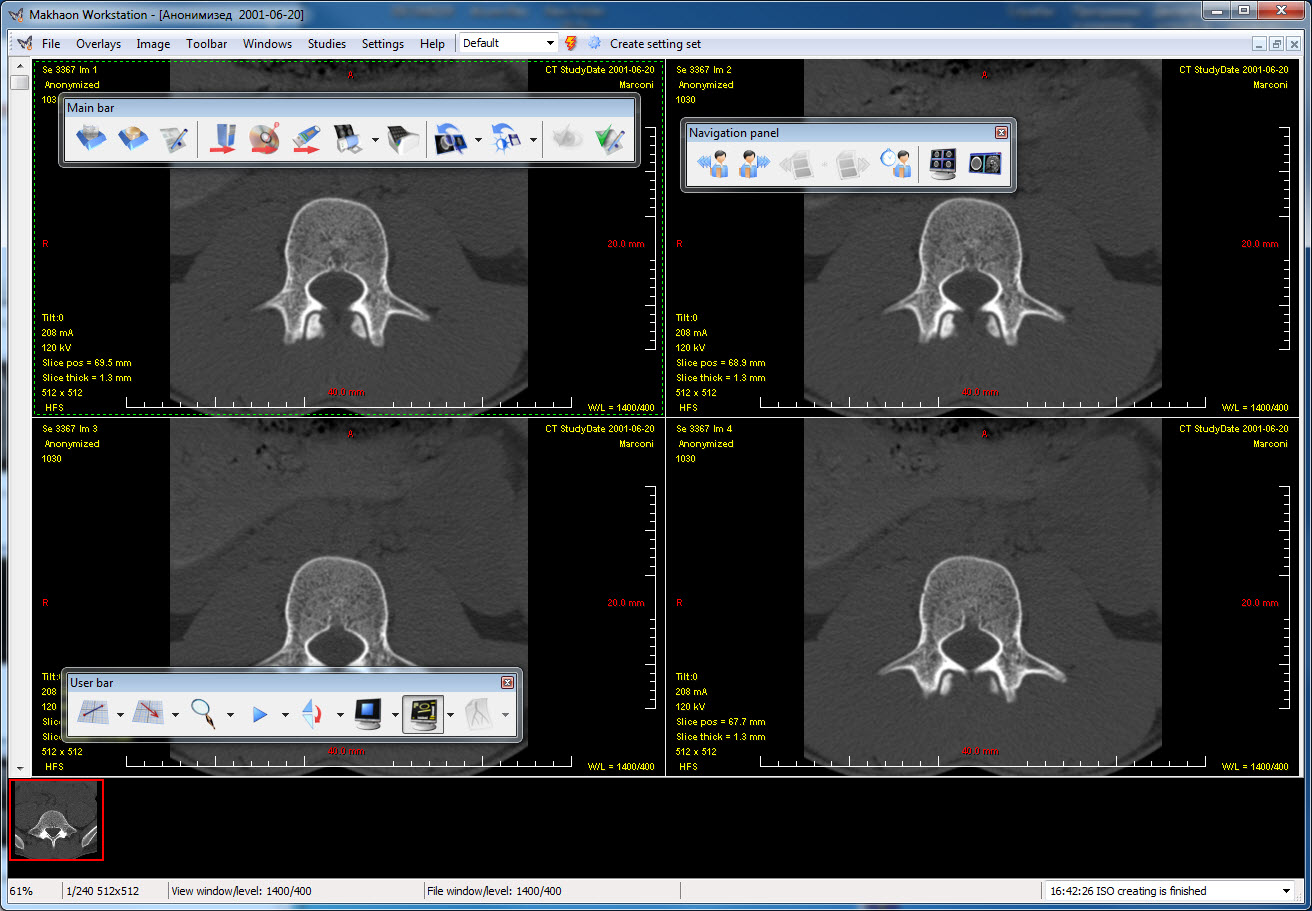
Main window. Controls position option.
Main window. Controls position option. Full screen mode.
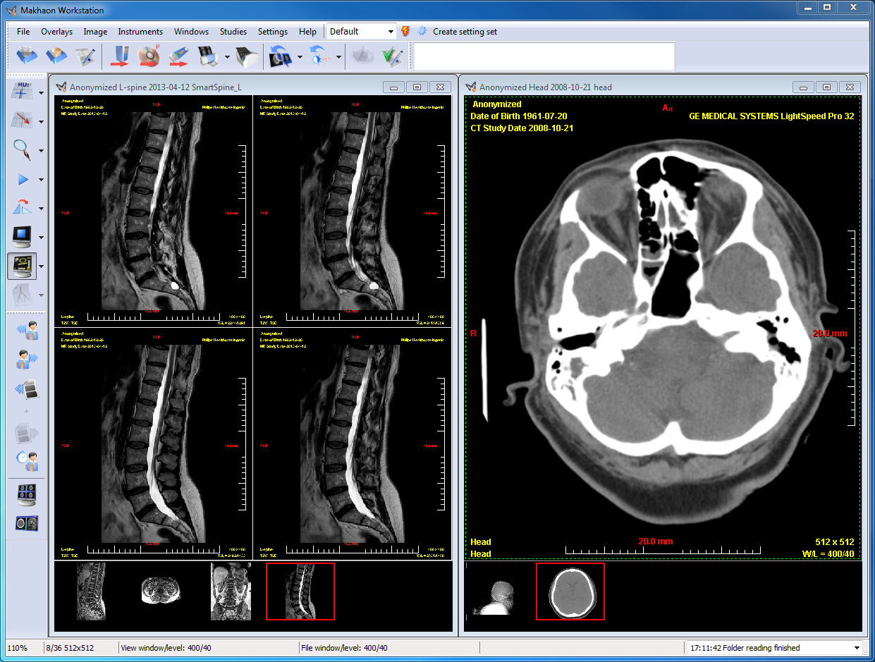
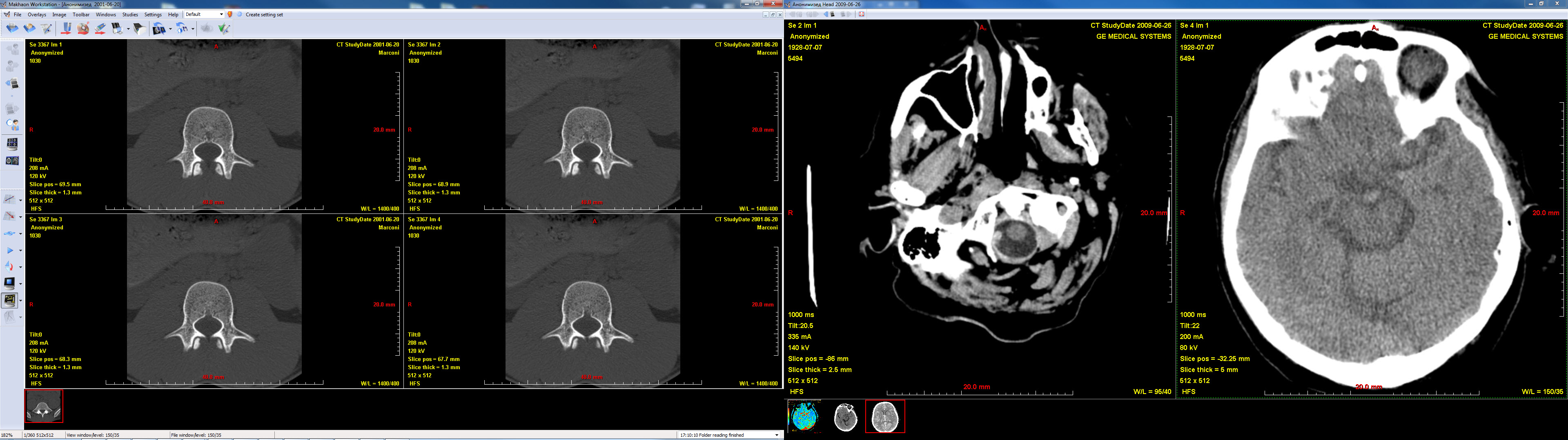
Main window. Work with two studies simultaneously. CT and MR studies are opened simultaneously, and different splitting is applied for different studies.
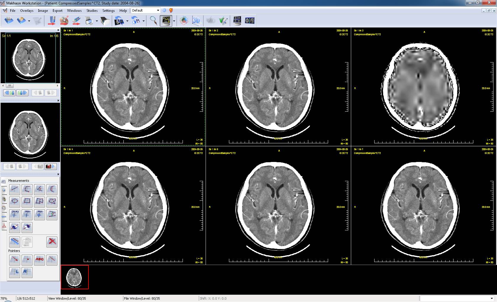
Series consisting of one and the same image, compressed in five different ways (plus one uncompressed image copy) is opened.
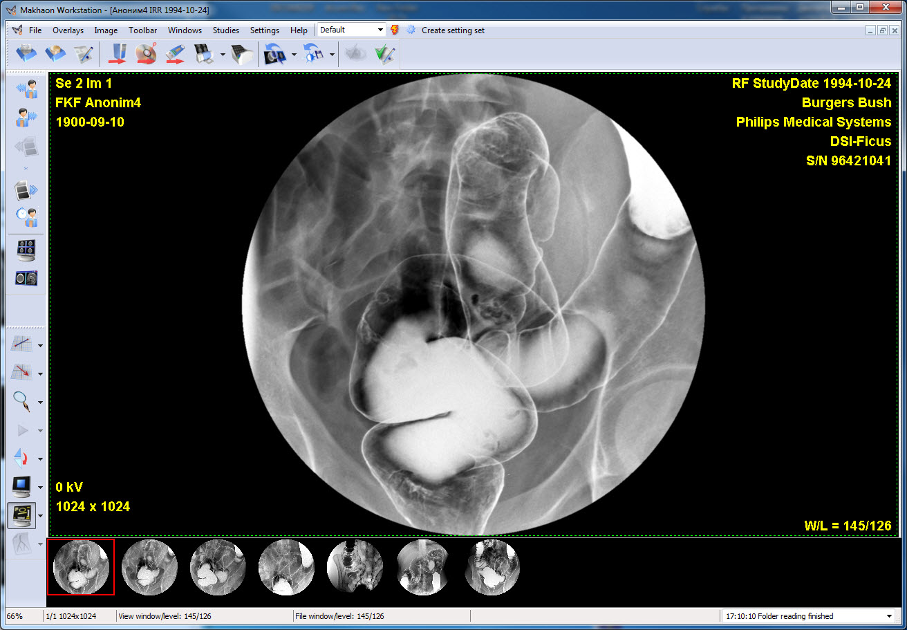
RF series - colonoscopy.
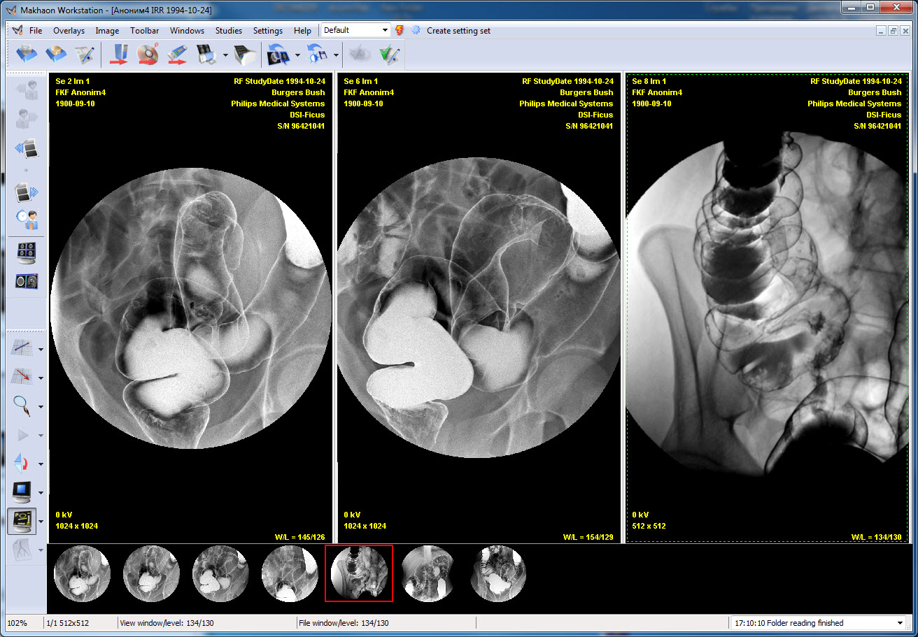
The colonoscopy. Three series are opened simultaneously.
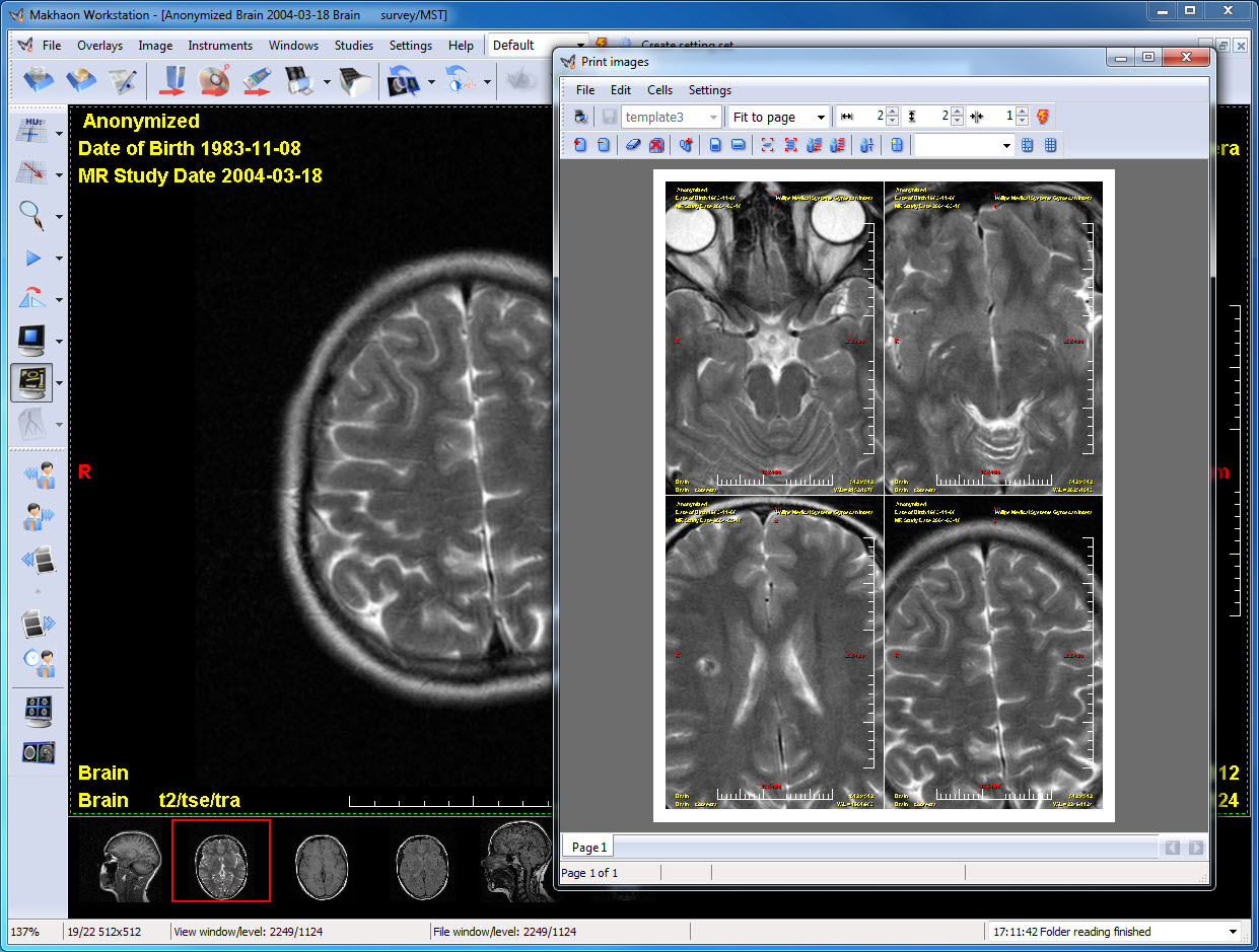
Image print window. Real size mode. In this mode image dimensions on the page or hard copy film are object actual dimensions.
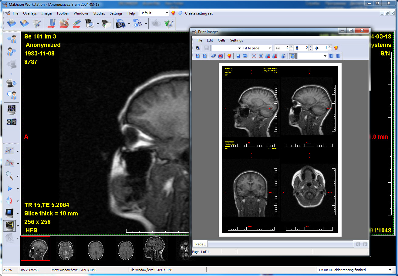
Image print window. "First image overlay" mode.
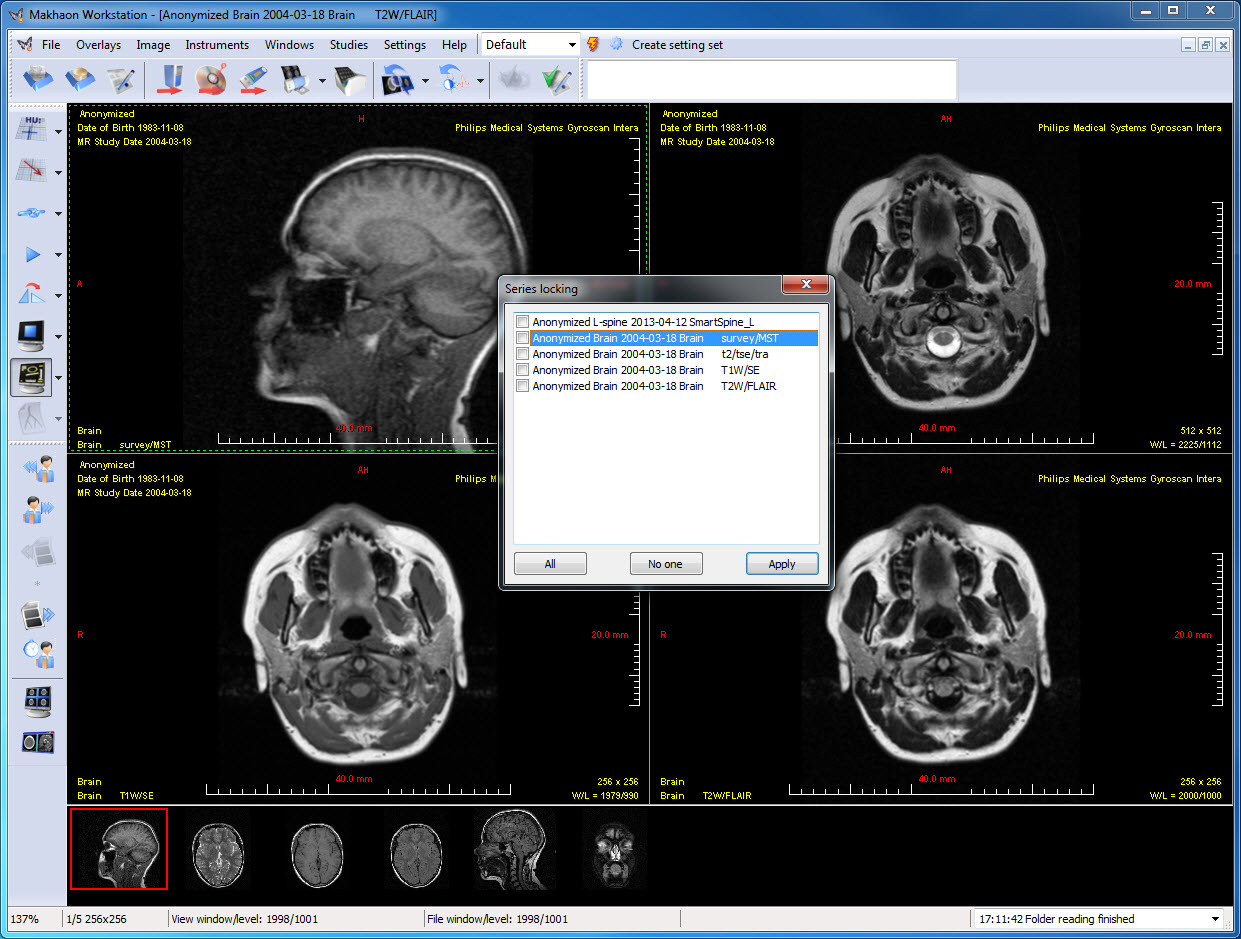
Any opened series synchronization.
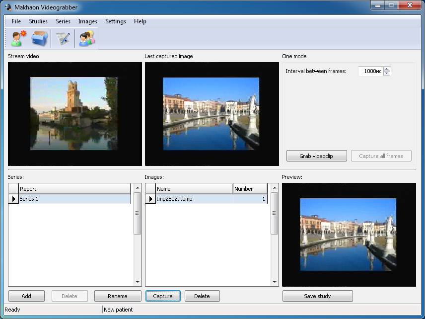
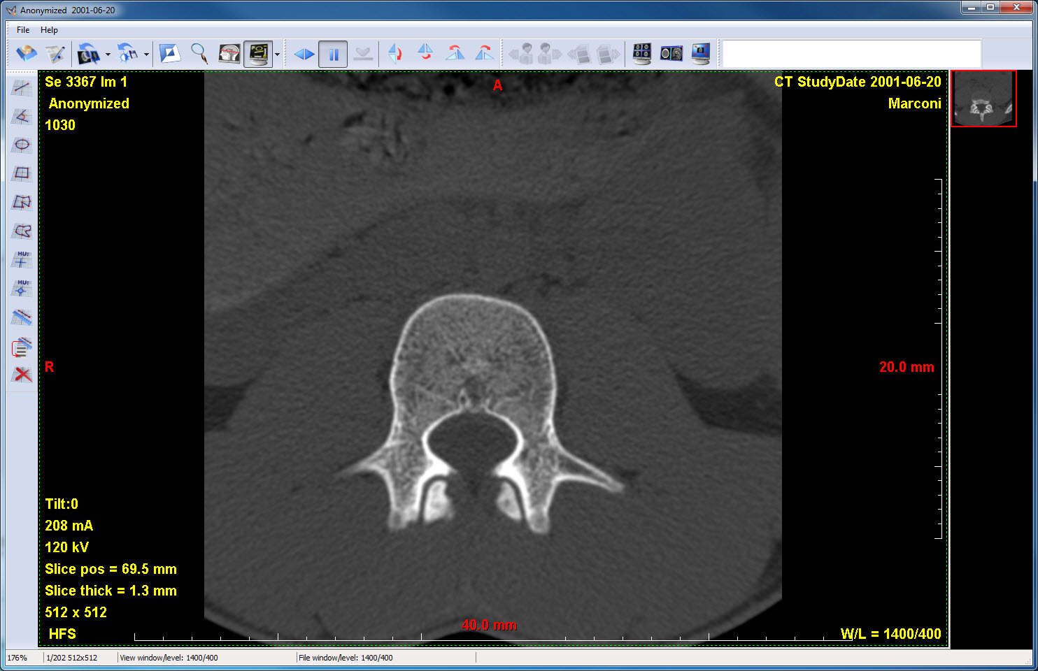
Video capture. Main window. One image is captured.
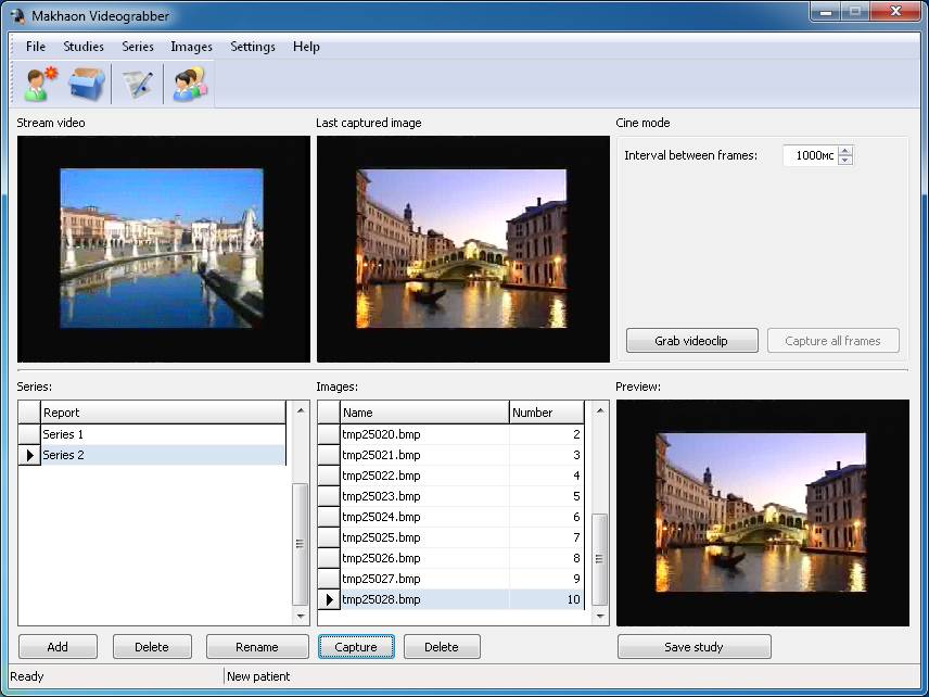
Video capture. Main window. Three image series are captured.
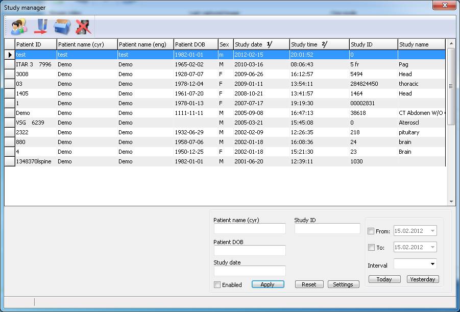
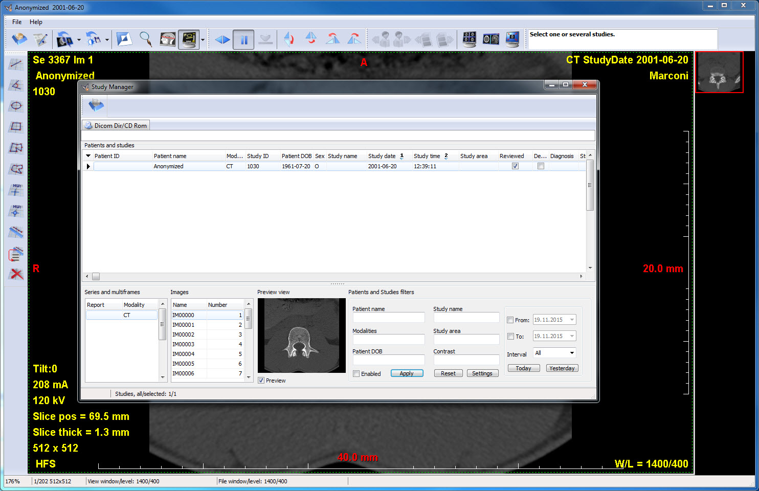
Video capture. Study manager. It is possible to make the new study, send existing ones to the DICOM device and add images to the studies.
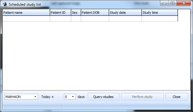
Video capture. Planned studies list window. If there is Worklist in the PACS system, then it is possible to enquire for the planned studies list. This functionality is operated on the DICOM Worklist protocol.
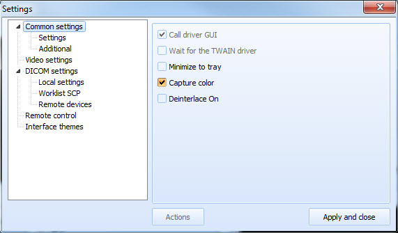
Video capture, options window. Tab "General settings".
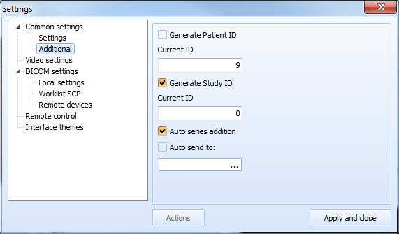
Video capture, options window. Tab "Additional".
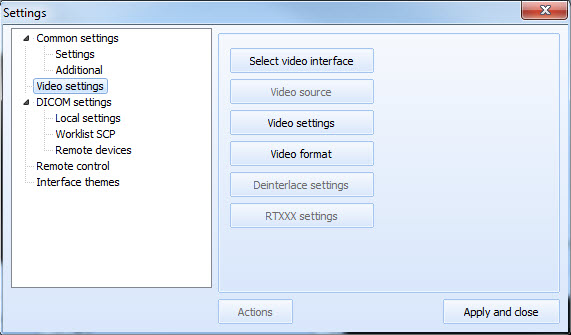
Video capture, options window. Tab "TWAIN and video settings".
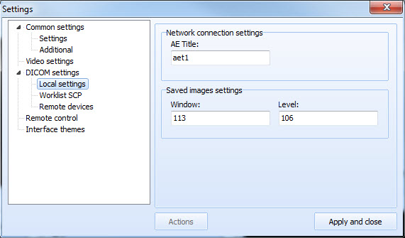
Video capture, options window. Tab "Local DICOM settings".
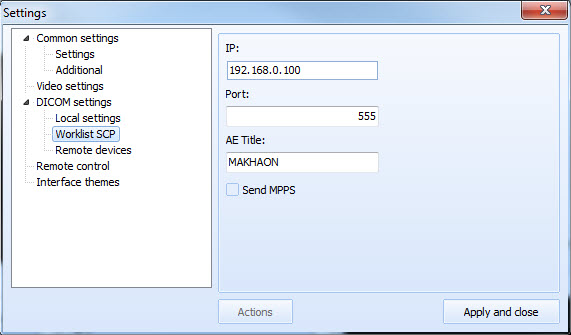
Video capture, options window. Tab "Worklist SCP".
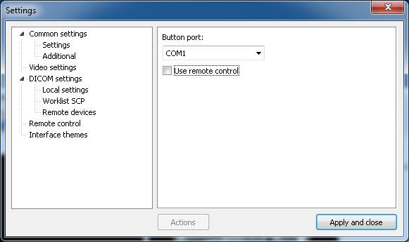
Video capture, options window. Tab "Remote control".
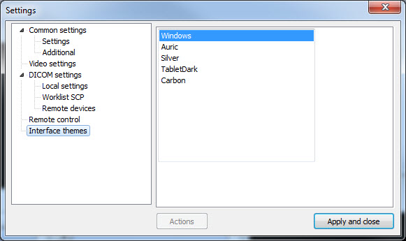
Video capture, options window. Tab "Interface theme". "Leopard" theme is applied.
Makhaon Lite. Main window. Makhaon Lite for CD and Makhaon Net Lite look almost the same.
Makhaon Lite for CD. Study manager. Net Lite has other tabs - local base and remote base.
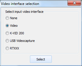
Video capture. Input interface option window.
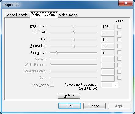
Video capture, options window. Video-source properties window.
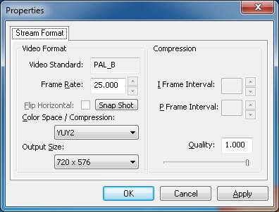
Video capture. Video stream properties window.
Simultaneous operation on two displays.
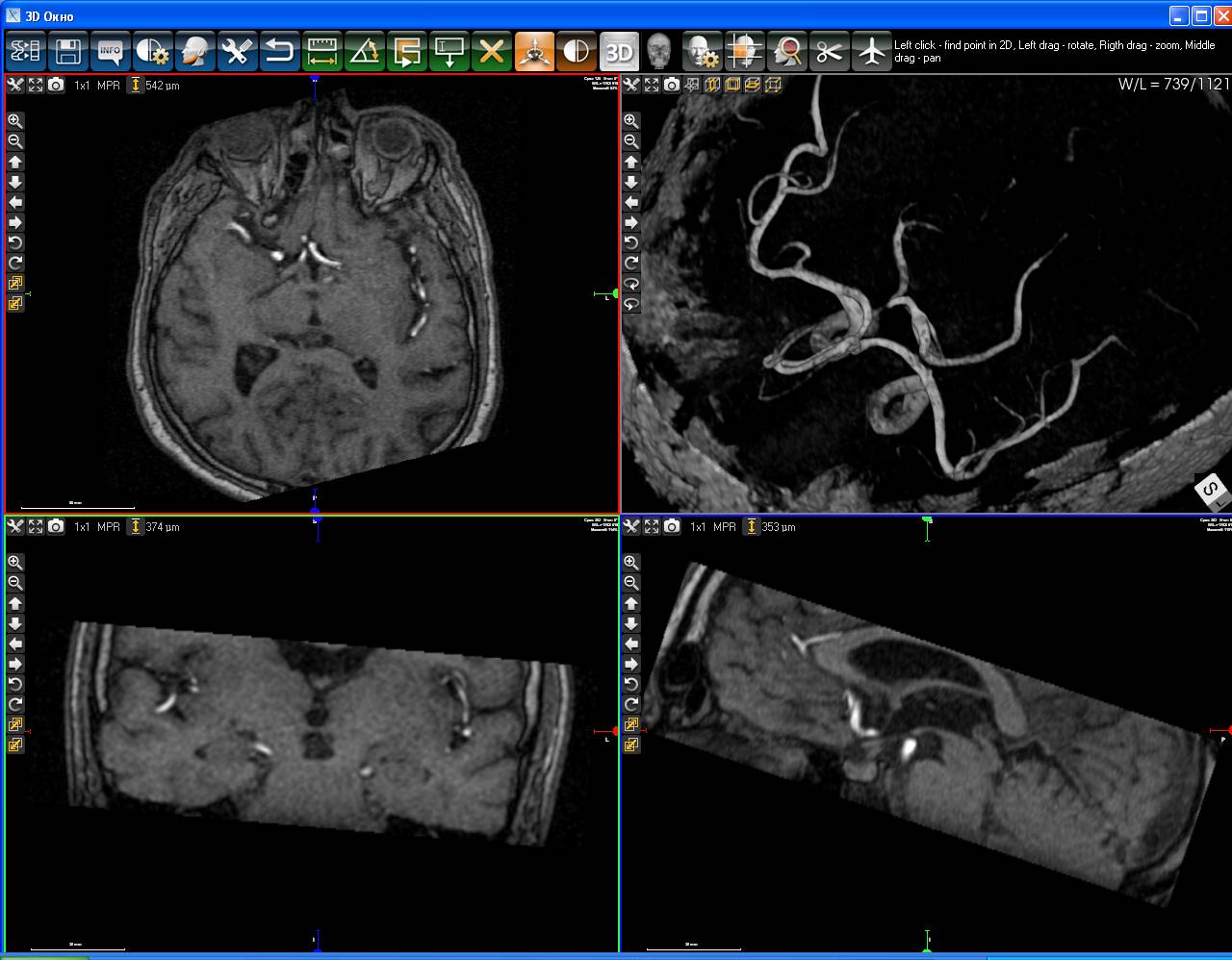
Magnetic resonance angiography.
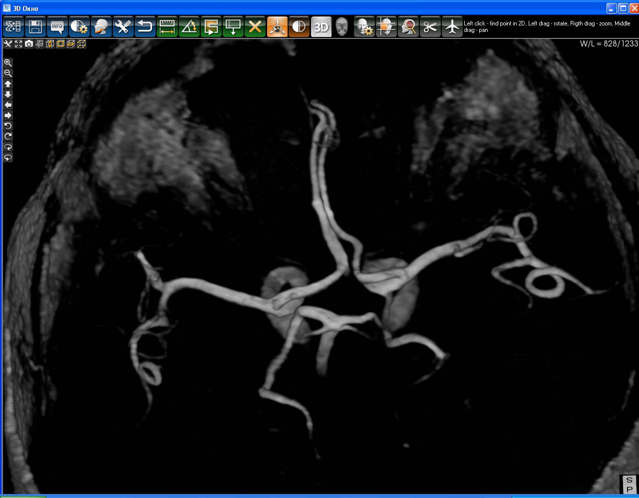
Magnetic resonance angiography.
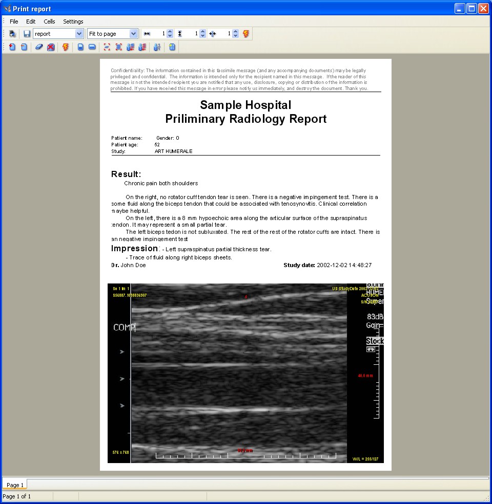
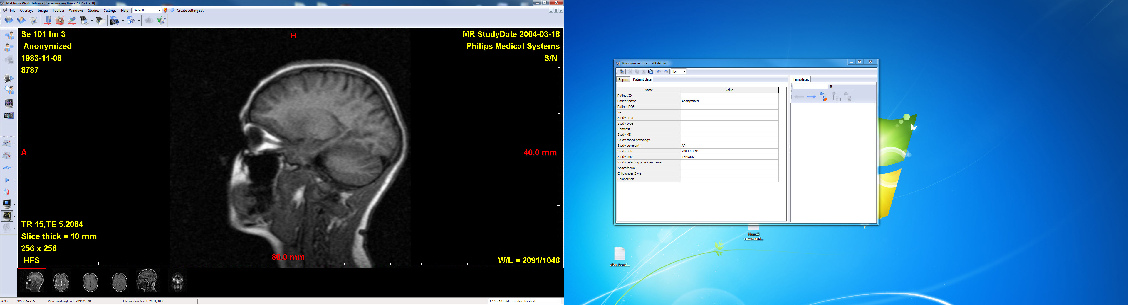
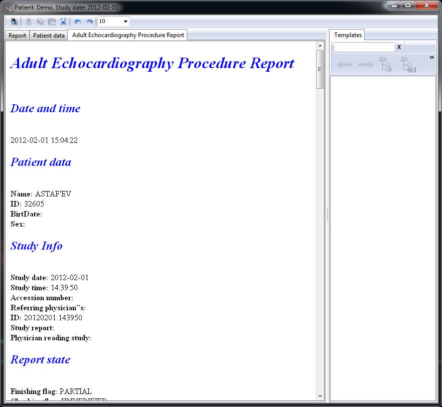
Study report with images.
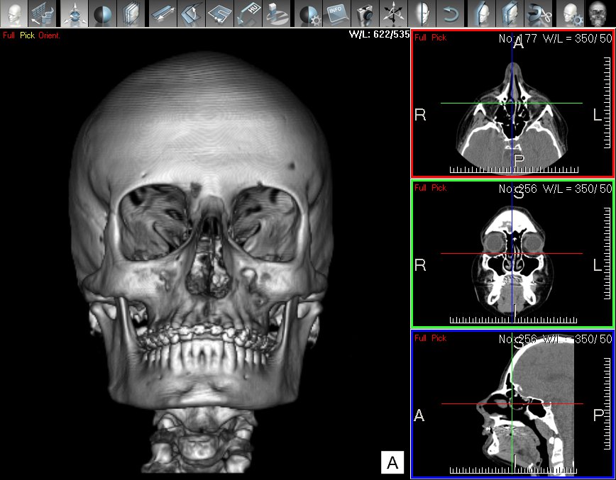
3D-visualization. CT-series.
3D-visualization. CT-series.
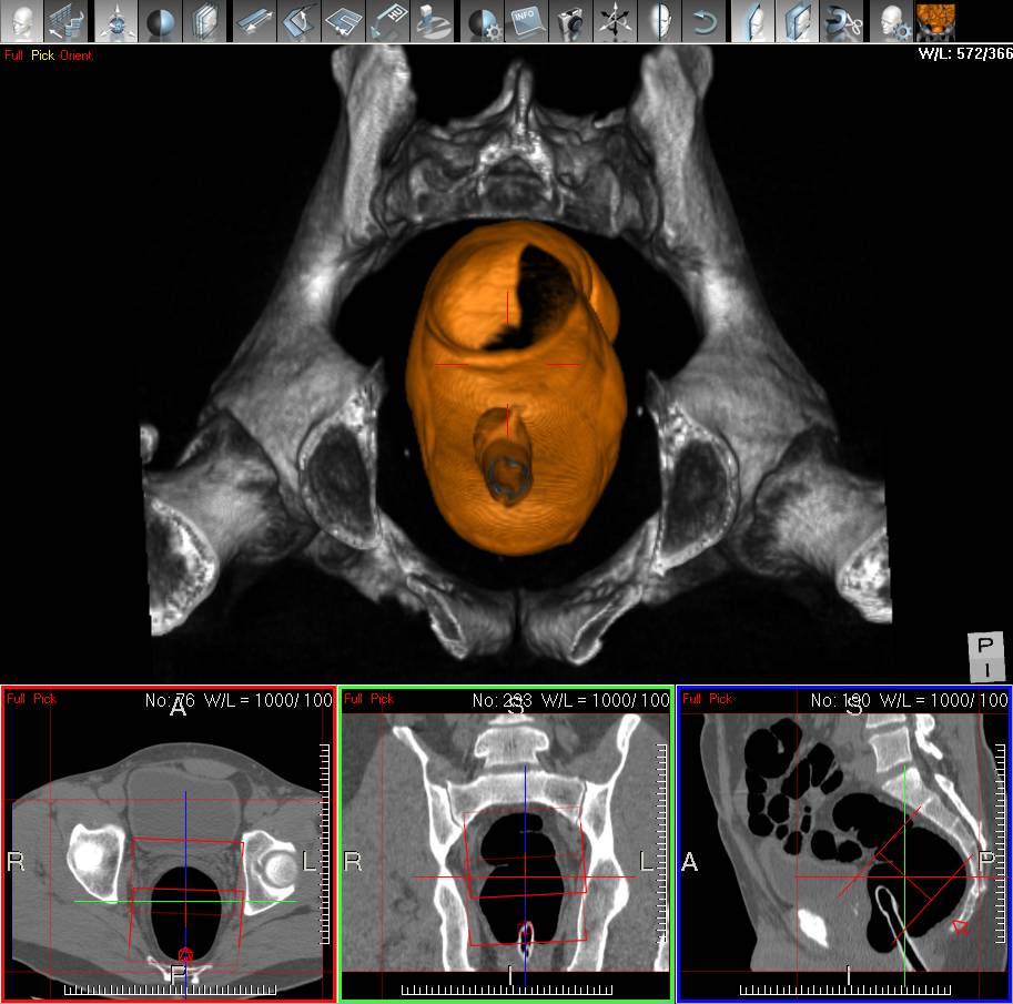
3D-visualization. CT-series.
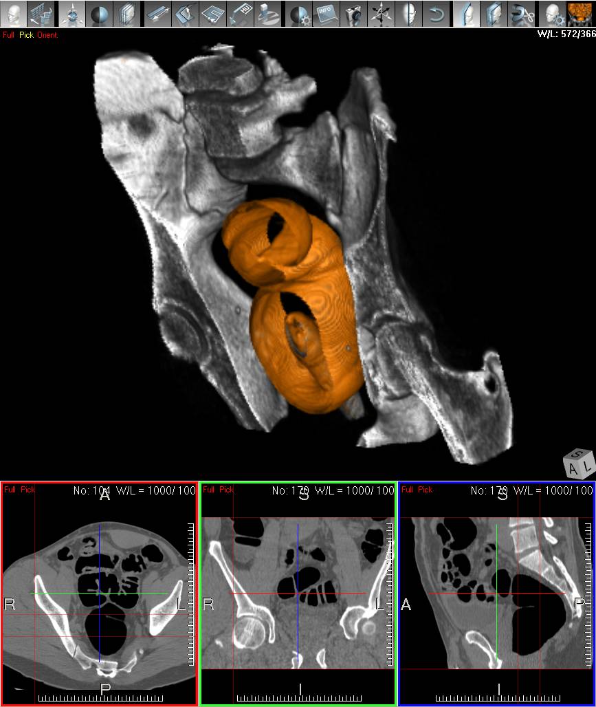
3D-visualization. CT-series.
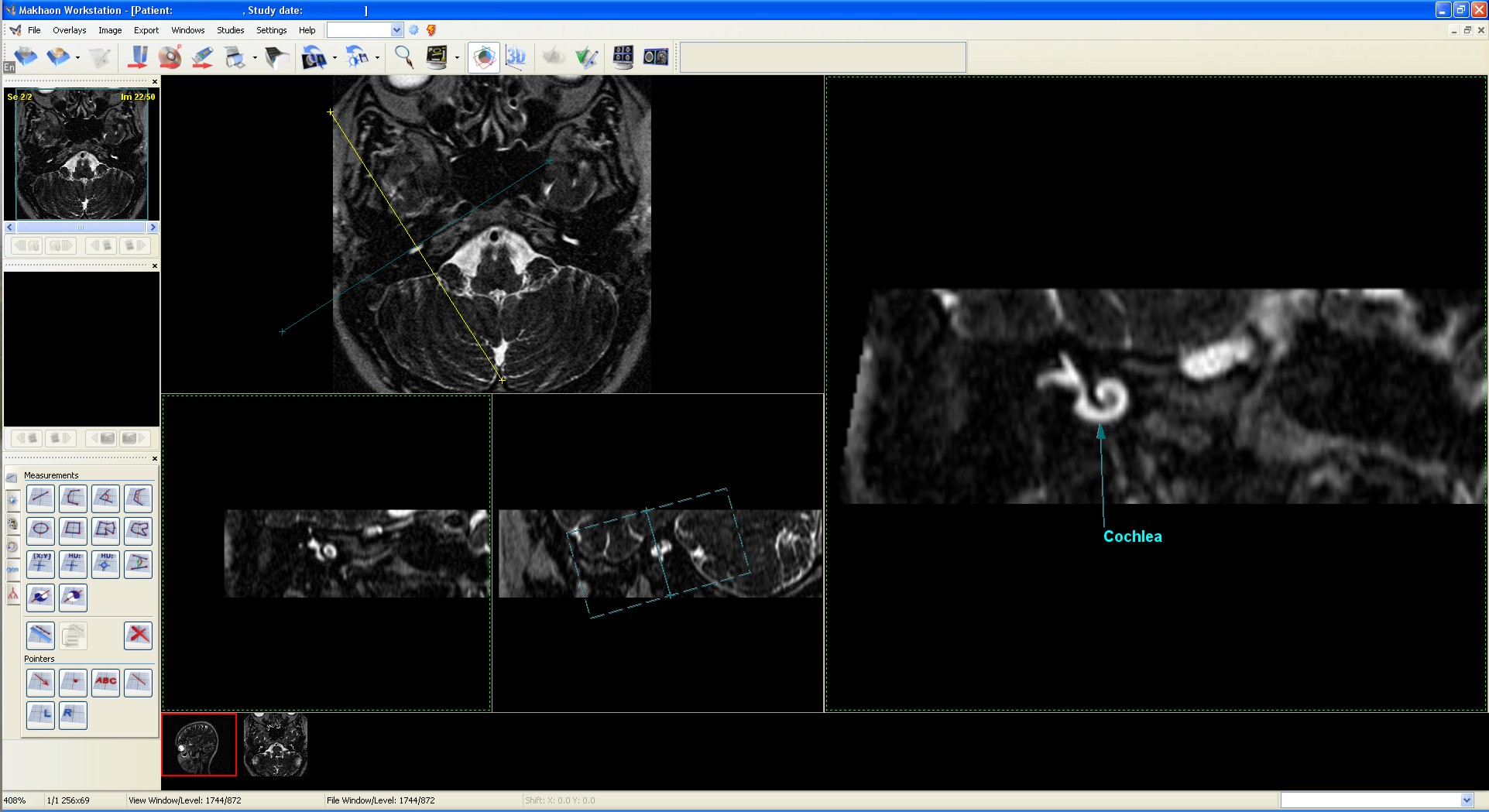
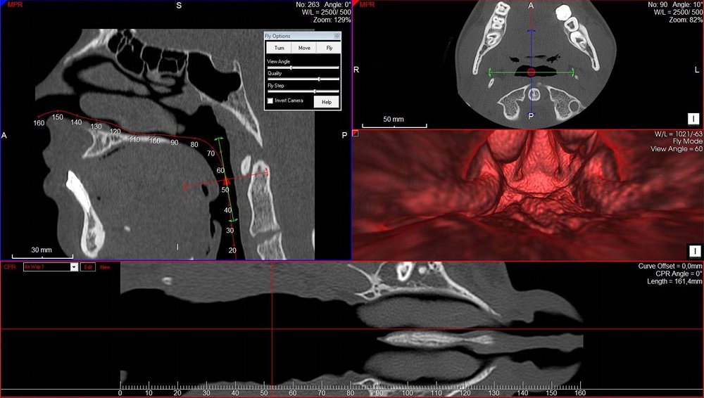
MPR usage in MR. Slice through cochlea.
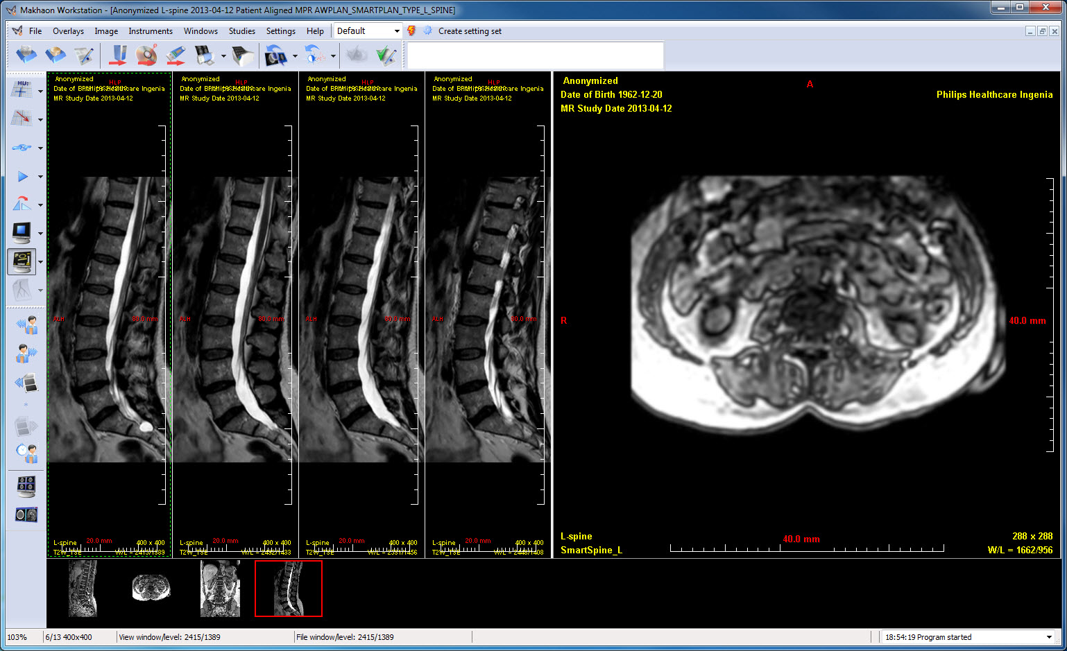
The MR series. The spine sections matched together.
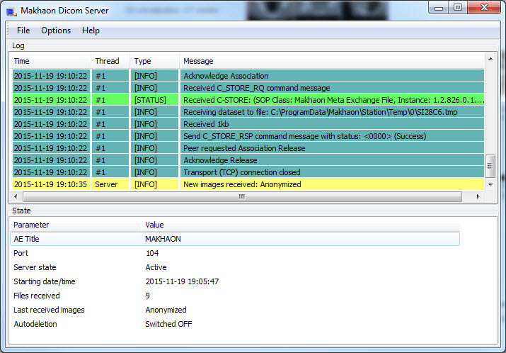
Makhaon server. MR image is stored.
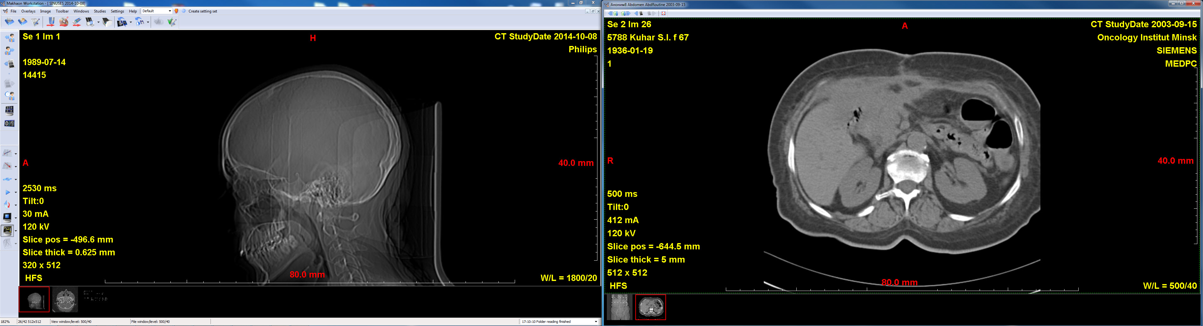
Simultaneous operation on two displays. Two CT studies are open.
Simultaneous operation on two displays. Two CT studies are open. On the left the 2x2-splitting is applied, on the right two series of study are displayed. It is possible to display studies on several displays simultaneously (up to 8 ones - that's the Windows restriction) using just one instrument "open on the free display". The station will find itself the display free from studies windows and will open the new window on it.
Structured reports created at US scanner Philips iU22. It is possible to review reports and print them.
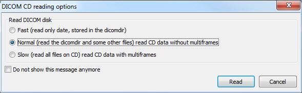
Dicomdir file read options window.