Site search
“Makhaon Workstation” package software
Makhaon Workstation features
Features Content:
- Common
- DICOM Support
- Graphic User Interface (GUI)
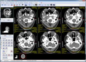
- 2D Viewer
- 2D view engine
- 3D viewer
- Full-featured study list
- Fast study list
- Studies import and export
- Working with CD (CD burning)
- Export to removable media
- Anonymization
- Advanced visualization tools
- Automatic settings (Hanging protocols)
- Study report
- Images and medical studies protocols print
- CD and DICOM archiving
- Multiuser mode, user authentication
- Integration
- Additional features
- Makhaon Workstation Benefits
Common
- Compatible with Windows XP, Windows Vista, Windows 7 and Windows 8 (32 and 64 bits OS)
- High performance native application. It runs in the native Windows environment, without using of .NET, Java etc.
- Fully parallel multi-threaded processing. Many processes are running in parallel to user interface mode for
maximize productivity
- Multi-language support in DICOM and user interface
DICOM Support
Images
- Multi-modality reading and viewing about all of existed DICOM files from all manufacturers (vendor neutral
view):
- Computed and Digital Radiography (CR, DR, DX, RF)
- Mammography (MG)
- Computed Tomography (CT)
- Magnetic Resonance (MRI, MR)
- Positron Emission Tomography (PET, PT)
- Ultrasonography (US)
- Digital Angiography (XA)
- Gamma Camera, Nuclear Medicine (NM)
- Secondary Capture and Scanned Images (SC)
- Intra-Oral Radiography (IO)
- Electrocardiogram (ECG)
- Hemodynamic Waveform (HD)
- Presentation State (PR)
- Encapsulated PDF (DOC)
- Structured Report Document (SR)
- Other Images (OT)
- Support single and multi-framed images
- Support XA, CT and MR multi-framed images (CT and MR enhanced images)
- Support RLE, JPEG Lossy, JPEG Lossless, JPEG-LS, JPEG 2000 Lossy, JPEG 2000 Lossless compressions
- Support MONOCHROME1, MONOCHROME2, RGB, YBR_FULL, PALETTE COLOR photometric interpretations
- Support 0 and 1 planar configurations
- Support 1, 2, 4, 8, 12, 16, 24 and 32 bits files
- Support explicit and implicit VR transfer syntaxes, little and big endian
- Support read of old DICOM files - which have no preamble or prefix
- Support standard DICOM grayscale rendering pipeline
- Support processing of the look-up tables (LUT). Modality LUT, VOI LUT and Presentation LUT can be processed.
The
8-, 12- and 16-bit gray-scale tables as well as 8- and 24-bit colored tables are processed
- Support processing the pixel spacing according to the X- and Y-axis (image calibration)
- Support 6000 group overlays
- Support bitmap overlays
- Support processing the color intensity, the orientation in space, slice thickness
- Support the US-regions processing. For the US images, not the integrated calibration of all the image but the
calibration of its separate areas is often used. For example, some areas can be calibrated in centimeters, the
other ones are in cm/sec. The calibration in dB, cm, sec, Hertz, dB/sec, cm/sec, cm/sec^2, cm^2, cm^2/sec,
cm^3,
cm^3/sec is also processed
- Support animation speed data processing
- Support angiography animation boundaries data processing
- Support dynamic mask processing in the DSA mode
- Support DICOM shuttering
- Support padding value processing on the CT images
- Support the Grayscale Softcopy Presentation States (GSPS) and Color Softcopy Presentation States (CSPS)
processing
(IHE Consistent Presentation of Images)
- The GSPS and CSPS intended for store information about overlays, window/level values, image rotation,
mirroring,
shuttering, look-up tables
- Support reading, saving, modifying, applying, exporting own and other vendor GSPS and CSPS (vendor neutral)
- The PSs can be saved in the database, be exported between the workstations and between the workstation and
DICOM
devices, can be recorded to the CD and read by the CD view program
- Support SR (structured report) processing. Structured report reading. The data contained in the SR can be
read,
displayed and printed on the printer (vendor neutral)
- Support encapsulated PDF reading, creating from a scratch and printing
- Support ECG and hemodynamic waveforms reading, rendering and printing (vendor neutral)
- Support specific ECG waves measurements: amplitude values in millivolts and in time in seconds
- Support hemodynamic measurements: pressure in mm Hg
- Support reading and displaying all DICOM tags
- Support of all standard DICOM character sets including Arabic, Cyrillic, Greek, Hebrew, Chinese, Japanese,
Korean,
Thai, unicode UTF-8
- Support reading, creating and modifying dicomdir
- Support view of non-DICOM images - JPG, BMP
Networking
- Support DICOM store SCU/SCP. Is a service meant for the images export to and import from the remote DICOM
systems
- Support DICOM query and retrieve SCU/SCP. Is a service intended for getting the list of studies from or to the
remote DICOM system and transfer the images to or from the other DICOM system
- Support DICOM print SCU. Is a service intended for printing the images on the DICOM printer
- Support DICOM storage commitment SCU. Is a service that lets you verify if files that were previously sent to
the PACS were stored by the application. Helpful for automatic archiving of the studies
- Support DICOM verify SCU/SCP (echo). Is the service created for checking the DICOM connection with the remote
DICOM system
- Support unlimited number of connected modalities and parallel connections
Graphic User Interface (GUI)
- Programs in package has simply, user-friendly, intuitive interface
- User interface constantly being improved to maximize productivity
- Support tool-bars customization, user can create own tool-bars or customize existed
- Support hotkey customization
- Support mouse binding customization
- Support wheel mouse
- Support easy mouse navigation, all operations like window-leveling, pan, zoom, scroll are handled with one hand
2D Viewer
- Support files opening without need for saving them into the database
- Support files opening with the help of "Drag and Drop" tool
- Support fast processing of big images (the images of 10 000 x 10 000 resolution were tested), the speed of any
operation performed over the image doesnt depend on its resolution
- Support work with big size images. This function was tested on the images of up to 2 GB in size (like the
multi-frame ultrasonic image for example)
- Support fast series opening. The series of 1000 images can be opened (and later be read) for 1-2 seconds
- Support series open on one or several sub-screens
- Support sub-screen layout split up to 8x8
- Support standard tool set ("zoom", "pan", "scroll", "win/level"), as well as many other tools
- Support fit zoom, 100% (pixel to pixel) or 200% zoom
- Support ROI (Regions Of Interest, Overlays): segment, polyline, angle, double angle, circle/ellipse, rectangle,
polygon, freehand, coordinate probe (2D/3D position display), HU probe, average HU probe, black gradient segment,
white gradient segment, arrow, point, text label, line, Letter 'L', Letter 'R', Cobbs angle, the "Screen
magnifier" and Region (trace contour, magic wand) tools
- Support shutter adding to the images series. The shutter can be rectangular or iris (octahedral), black or white
- All overlays are subdivided into 3 groups: "DICOM overlays", "Positioning overlays", "Measurement overlays". User
can switch them on or switch them off as each one separately, as all together
- Support user defining color, size and fonts of all overlays
- Support font autosize mode. Size of overlays is changing when changing workspace size
- The overlays state will be taken into consideration in case you add any images to the print manager
- Support add, edit, delete, copy, past, save (to presentation states), restore (from presentation states), send to
network, receive from network, write to CD, read from CD measurement and positioning overlays
- Support measurements of distance, angle
- Distance and angle measurements show current value during drawing
- Support length measurement using estimated radiographic magnification factor (for MG images)
- Support measurements of square, perimeter, average hounsfield units (or signal intensity) of the areal overlays
- Distance measurements can be made either in mm, in micron or in nm (the two latter ones are more appropriate for
microscopy), the calibration is taken from the DICOM files or can be created by user
- Support ruler display
- Support images orientation label
- Support image orientation changing while doing flip and rotate
- Support image rotation clockwise or anticlockwise at an angle of 90 degrees, image mirror relative to the
horizontal and vertical axis. This functions can be applied to a single image or for a whole series
- Support image analysis with help of a image histogram calculation
- Two different histogram can be shown. Histogram of the linear measurements looks like a curved line and histogram
of the areal measurements looks like a bar histogram
- Support statistic parameters calculation for a current histogram: minimum, maximum, population variance, standard
deviation, mean value, euclidean L2 norm
- Coordinate grid on the histogram is calibrated in mm and HU (in case an image calibration exists)
- Support "reverse order" mode. The opportunity to view the images from the last and up to the first one (on default
it is the images view from the first one and up to the last one)
- Support to open several series simultaneously. The number of series is restricted by the RAM size
- Support simultaneous opening of several series of one medical study
- Support simultaneous opening of several series of different studies
- Support opening of study (studies) on different screens simultaneously. The number of them is restricted by the
RAM size
- Can show series thumbnails. Thumbnails are created from the middle images of every series of every opened medical
study
- The series thumbnails are loaded in the separate thread and not to slow main series view process
- Thumbnails can be placed on any side of workspace or can be disabled
- Support the "Current image" mode - the image specified is displayed in full-screen size, the full-screen mode exit
recover the screen subdivision and the specified image display
- Support fully customizable display tags from a DICOM file
- Tags can be shown in any image corner, many tags per one line
- Explained strings can be added to tag values, station can show tags like this: 'Slice thickness 5.0 spaces between
slices 3.0'. Any strings can be mixed with any tags (and any numbers of tags) values
- Tags values smartly rounded for readable view: 3.99999 shows like 4.0 etc.
- Support digital image filters; 3õ3 convolution filters: The Laplace, Hipass, Find Edges(Top Down), Sharpen, Edge
Enhance, Color Emboss, Soften, Blur, Soften (Less), Lap 1, Lap 4, HP3, Find Edges (Bottom Up), Sobel Hor, Sobel
Vert, Previt Hor, Previt Vert, Dilatation, 7 bit, Median
- Support fast filter reset function performed by one keystroke
- Support inversion mode. In most cases there are no need to change the window/level values after inversion -
window/level values are automatically calculated for the inversion mode
- Support different series or studies compare
- Support synchronized view between two or more series
- Support animated displaying of the series images
- Support animation in "First frame to last" and "Play forward and backward" modes
- Can animate with 60-80 frames per second speed
- Support manual animation speed adjustment
- Support for real-time mode (the speed being automatically calculated from the DICOM data)
- Support three scrolling modes: the "Standard mode", "All images simultaneously", "Page-by-page"
- Support the use of window/level preset values (mostly for CT - chest, head, lung, bone etc)
- Support binding preset values to the keyboard buttons
- Support adding, editing and delete preset values
- Support user defined look-up tables (LUT) (pseudo color mode)
- Support colored and black and white (brightness levels) LUT
- Support linear and non-linear LUT
- Support LUT creating, editing, saving and deletion
- Support automatic window-level calculation
2D view engine
- Super fast 2D view engine with usage of 3D Direct Draw hardware accelerator
- Support hardware and software rendering
- Works fine on all, even old and integrated graphic cards
- Support bi-cubic, bilinear and nearest neighbor interpolation in software and hardware (with usage of pixel
shader) modes
- Support multi-monitors systems, up to 8 monitors, including different resolutions and color depths
3D viewer
- Support Multiplanar Reconstruction (MPR)
- Support thick slab calculation (raysum, MIP, MinIP, scout image)
- Support change slice thinkness
- Support curved MPR (CPR)
- Support 3D Maximum Intensity Projection (MIP)
- Support 3D volume rendering
- Support 3D surface rendering
- Support 3D measurements
- Support export full-functionally (16 bit, normally window-leveled, with full spatial information) series of
reconstructed images
- Support 2D and 3D fusion mode for view fused PET-CT series
- Support bone removal
- Support 3D implant fitting
- Support special dental mode
- Support volume calculation
- Support 3D templates editing
- Support 3D fly mode
- Support virtual colonoscopy or endoscopy
Full-featured study list
- Can store unlimited number of images in the local data base
- Can store instances in the separated files (not in data base), stores files as is, without modifications, creates
local Vendor Neutral Archive (VNA)
- Can show next lists: local data base; remote data base (result of DICOM query); structured DICOM CD-ROM
(dicomdir); non-structured DICOM folder; CD archive; temporary data base
- Can sort list on any fields within the database, descending and ascending
- Can filter list by any six fields simultaneously
- Support additional filter by range of study dates, by date interval
- Support fast search filters today, yesterday
- Can preview image on studies, series and images from any data base (from remote data base with help of private
tags from Makhaon Storage)
- Support medical studies info saving in DOCX, XLSX, HTML, TXT, CSV, XML formats
- Support import and export medical studies from and to different sources
-
Support 'on demand' capability. Can open studies directly from a CD or a folder without downloading them into the
local database first. Can open studies from a DICOM server immediately after downloading
Fast study list
- Can access to the studies from last two days in local database
- Support fast on-the-fly filter by patient name
- Support open study for view in current workspace, in new workspace, create workspace on free monitor, different
series on different monitors
- Support list of prior studies found in local base, remote DICOM base, CD archive
- Support image preview of selected studies in local and remote base
Studies import and export
- Support import from a remote base (with the help of DICOM Retrieve service), vendor neutral
- Support import from a compact disk or any directory structured as standard DICOM media (that contains dicomdir
file), vendor neutral
- Support import from non-structured local or network folder
- Support direct import DICOM files from a local or network folder to the local database
- Support import from non-DICOM files (dicomization); from JPG, BMP or PDF files
- Support export studies to DICOM, to the non-medical graphic formats (JPG, BMP, TIFF) or to the cine-loops
(AVI)
- In case of AVI format image export the images additional compression with the help of codecs installed in Windows,
the frame width and height setting, the image compression degree setting, the replay speed setting and the codec
parameters setting are possible
- In case of image export to AVI the replay speed is taken from DICOM data (if the corresponding data is available
there)
- Can save AVI files separated by the series. It happens in following way: the user selects one or several studies
(one or several series), the program saves the selected studies or series separating them in several AVI files.
One AVI file is created for one series. All usual AVI options as the frame size, codec, image quality,
compression, etc. are available here
- Support export studies to CD (CD burning)
- Support export studies to removable media (to the flash)
- Support export studies to DICOM network (DICOM Store)
- Can do fast copy of the current image to clipboard or save it in DICOM, JPG, BMP or TIFF format
Working with CD (CD burning)
- Can select the CD burner from list
- Can select burn speed of device
- Can show compact disc and recorder information
- Can erase information from the rewritable discs (fast or full modes)
- Support recording the information on the CD-R, Double density CD-R, CD-RW, Double density CD-RW, DVD-R, DVD-RW,
DVD-RAM, DVD+R, DVD+RW, Dual Layer DVD+R, DVD-R, Blu-Ray Recordable, Blu-Ray Recordable & Erasable, Dual Layer
Blu-Ray Recordable & Erasable, Dual Layer Blu-Ray Recordable, HD-DVD Recordable, HD-DVD Recordable & Erasable,
Dual Layer HD-DVD Recordable & Erasable, Dual Layer HD-DVD Recordable
- Support recording of the compact discs corresponding to the part 10 DICOM (part 10, p. 10) with view program or
without it (station can works like a File-set Creator, FSC)
- Support DICOM conforming DICOMDIR creation
- Can add lite version of workstation to CD (portable Makhaon Lite)
- Support JPG compact discs recording
- Support creation of HTML pages, structured according to the patients names, medical studies and series on JPG
discs (for Android, iOS, MacOS, Linux, Unix Users)
- Support combined compact discs record (JPG and DICOM simultaneously)
- Support medical study report record to the CD
- Support medical studies addition to the already recorded discs created as with the help of the "Ìàkhaon
Workstation" software itself as with the help of other software able to work with the standard DICOM p10 media. In
this case the DICOMDIR files, HTML, JPG files structure and medical studies reports structures are automatically
updated (station can works like a File-set Updater, FSU)
- Support verification of burned data
- Support archive disc recording
- Can create ISO image
- Support of anonymization
- Can show detailed log while CD created and burned
- Support burning on-the-fly, without DICOM files copying to the temporary folder and without temporary disc image
creation. In this case the recording process takes considerably (up to two times) less time and there isnt any
need for much hard drive space
- Can fast burn any opened study from the Desktop window of the application
Export to removable media
- Support removable media selection
- Support automatic update of removable media list while plug and unplug removable devices
- Support safe device ejection
- Can show detailed log during export process
- Support all export features like to the CD - DICOM part 10 export, JPG export (with HTML creation), medical
studies reports export, update information (File-set Updater)
- Support export to a separated folder (not only to root)
Anonymization
- Support anonymization while export DICOM files
- Support anonymization while send studies to DICOM node
- Support anonymization while CD burned
- Support anonymization while export to flash
Advanced visualization tools
Angiography
- Support digital subtraction for angiography (DSA)
- In the subtraction mode all the usual modes and techniques applied to the usual images are possible:
measurements,
digital filters, color schemes, diaphragms, print, real-time animation, and so on
- Support fast, real-time algorithm (with MMX usage) for the digital subtraction. The speed in DSA animation
mode is
up to 60 frames per second
- Support subtraction of 8-bit and 16-bit images
- Support dynamical masks switching in the static and in the animation mode. Information about mask applied to a
current image is taken from DICOM headers
- Support real-time WYSIWYG mask shifting
- Support mask shifting by fractional pixels (by 1/10 of pixel) for the best overlaying
- Support mask shifting for the whole series or for the single images only
- Support changing parameters of subtraction (subtraction level and subtraction factor)
- Support mask averaging from images
- Support calibration by catheter
Tomography
- Support localizer line (scout image, scout line, scanogram, reference line) on the MR and CT images
- Support localizer line calculation on any intersecting images
- The localizer line takes account of the image size and indicates correctly the positional relationship of the
images
- Support MPR (multi-planar reconstruction) with orthogonal or double oblique image planes
- Support compound series (such as multi-phase, multi-component and simply pasted ones) automatic subdivision
with
fast selection of subdivided series. The algorithm of automatic splitting was tested on the great number of
various
compound series
- Support automatic rejection of images inadequate for reconstruction (for example the ones without angular
points and
directing vectors)
- Support fast, real-time, reconstruction even in the double oblique mode
- Support reconstruction for 8-bit and 16-bit images
- Support image fusion for PET-CT studies with adjustable blending percentage
- Support 3D cursor mode on regular series and series with oblique slices
Mammography
- Support background image air suppression while image window-leveling or inverse
- Support displaying of the overlays and marks, received from the automatic diagnostics programs (display of CAD
SR)
- Support automatic layout of the images - RCC, LCC, RMLO, LMLO are merged from different series to one, sorted
and
mirrored (if needed)
- Support automatic mammo image view mode. Station split workplace to 2x1. If system has two or more monitors,
station
automatically shows second workspace window on second (or other, monitors to show images can be customized)
monitor
- Support 1x1, 2x1, 2x2 and 4x1 workplace splitting
- Support pairs-linked mirrored (from center of the workspace) images pan and zoom
- Support pairs-linked image rotation and mirroring
- Support omitting of 'for processing' (nonscreening) images
- The screen overlays is displayed at the opposite side of the breast to avoid being overlapped with the
mammography
images
- Support fit images to workspace to show it in same size
Other
- Support volume calculation
- Support density ratio calculation
Automatic settings (Hanging protocols)
- Support hanging protocols matching by modality, study name and study area
- Support hanging protocols creation, editing, applying (during or after series open), saving and deleting
- Support automatic screen laying out
- Support automatic zoom
- Support automatic image calibration
- Support selection of monitors to view the images
- Support automatic color scheme switching
- Support automatic reverse order applying
Study report
- Support description of medical studies including medical opinion on the study with the opportunity to use the
predefined templates
- Support export reports to other Makhaon Workstation or Makhaon PACS
- Support templates saving in the form of a tree with the opportunity for editing, adding or delete the brunches
- Support possibility to add, change or delete the phrases in the templates
- Support separate templates binding to the different fields (that is to the description, medical opinion or every
personal information fields)
- Support export medical studies results into the HTML format
- Support export results to clipboard
- Support export results to Windows printer (print a protocol, create paper hard copy)
- Support change patient and study information (patient name, patient id, study id, etc)
- Support words coloring. It's helpful for fast change of words in text like a 'left' to 'right', 'multiple' to
'single' etc.
Images and medical studies protocols print
- Support study results print template changing (template is in HTML format)
- Can print medical reports with hospital information and hospital logo and any other needed information
- Support addition any information from the data base to report (patient name, patient id, study date etc)
- Support printing of study protocol and images on Windows printer
- Support printing of images on the DICOM printers
- Support gray scale and color DICOM printers
- Support DICOM print on the paper, clear film, blue film, clear mammo film, blue mammo film
- Support the choice of Windows and DICOM printers and their parameters setting
- Support WYSIWYG preview before printing (print composer)
- Support multi-page print composition
- Support fully customizable print pages
- Support merge cells of printing workspace. Can freely add merged cells, and reset merging
- Support page parameters setting (borders, page orientation for Windows and DICOM printers)
- Support change print page laying out to up to 10X10 cells
- Support adding background to text overlays
- Support user defining color, size and font of printing overlays
- Support adding image to the layout from one or from different studies and from the scanogramm window
- Support automatic adding of all the current series to print. The program automatically creates the necessary print
pages
- Support change window/level, shift and zoom parameters. The parameters can be changed either for every separate
image or for all of the images on the page at once
- Support measurements adding or editing right on the print manager
- Support dragging the image in the layout from one cell into another
- Support set the real size of the images. The film copy image size corresponds to the physical size with the
minimum
error (mostly due to the errors in the film sizes)
- Support drawing the localizer lines on the selectable image(s)
- Can assign any image(s) to any other for drawing the localizer lines
- Can select all images of the series with one keystroke (for simplify further assigning)
- Support (switchable) draw of small scout images in any corner of main image
- Support the "Overlays in first cell only" mode. This mode can be useful for reducing the amount of information on
the images
CD and DICOM archiving
- Support archiving studies on CD or on DICOM nodes
- Support automatic selection of studies for archiving on CD. The studies are chosen with regard of the space on the
disc available, and then they can be recorded to the archive disc
- Every archive disc gets the unique number (within the local studies database) according to which it can be
identified afterward
- The number of the storage discs is restricted only by the local database file size (medical.gdb)
- The study on archive CD can be copied or viewed right from the disc. The program will suggest you to insert a disc
and will give its number
- The archive discs are the discs in DICOM p10 format and they can be read on the side working stations (vendor
neutral)
- It is possible to archiving on remote DICOM nodes. In this case storage commitment request is send to the node. If
node supports storage commitment SCP, it response to the program when it successfully stores instances it its data
base. After response Makhaon Workstation can automatically delete the archived studies if it is configured to do
it
Multiuser mode, user authentication
- Support the possibility to have several users in the Ìàkhaon Workstation package (users adding and users delete
is
performed with the help of "Ìàkhaon Configurator" program, being the part of the package)
- Support restricting access to medical informations
- Support hashed password protection of all the users records
- If there is at least one user in the package then Ìàkhaon Workstation is switched to the user authentication mode
and will require the users name and password for each program opening
- Support bounding users settings of the program interface, templates, window/level presets and other program
options
to every user
Integration
- Support OLE and command-line automation to open studies from the local database or from remote database
Additional features
- Support automatic retrieval prior studies from DICOM network, local data base or local CD archive
- Support search of priors by patient name, patient id, or fuzzy phonetically search
- Support fast view of prior studies
- Support full screen display
- Support plug-ins system, that can arbitrarily modify instances before storing it to data base
- Support archived study auto-deletion
- Support view DICOM header
- For increase performance some parts of software (all window-leveling, parts of LUT engine, DSA, convolution
filters)
are realized in regular or MMX assembler
- Support data base backup and restore
- Support change location database of images
Makhaon Workstation Benefits
- Support receive, send, open, store, work with images from different vendors (vendor neutral)
- Supports all common used modalities in one software
- Designed as failsafe routine everyday tool
- Easy to learn and use. Software usage usually does not require specific training
- Fast and responsible software support, users' forum, internal bug and feature tracking system, continuous internal
beta-testing
Products
Screenshots
Download demo